

Search results: LOX CRISPR
Product group Assays
Assay Sample TypeSerum, plasma and other biological fluids.
ReactivityBovine
- SizePrice
Product group Assays
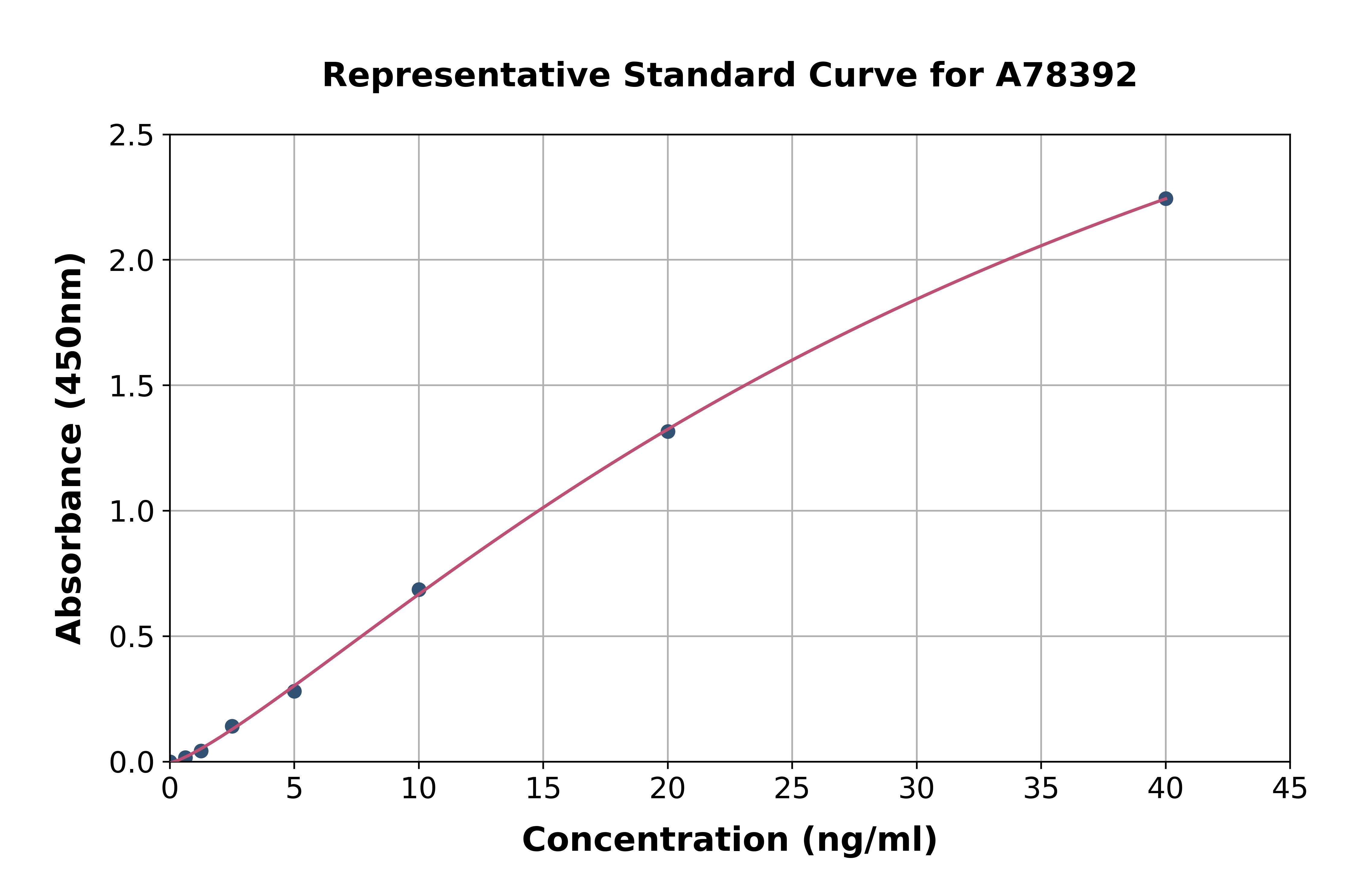
Human LOX ELISA KitA78392
Assay Sample TypePlasma, tissue homogenates and other biological fluids.
ReactivityHuman
- SizePrice
Product group Assays
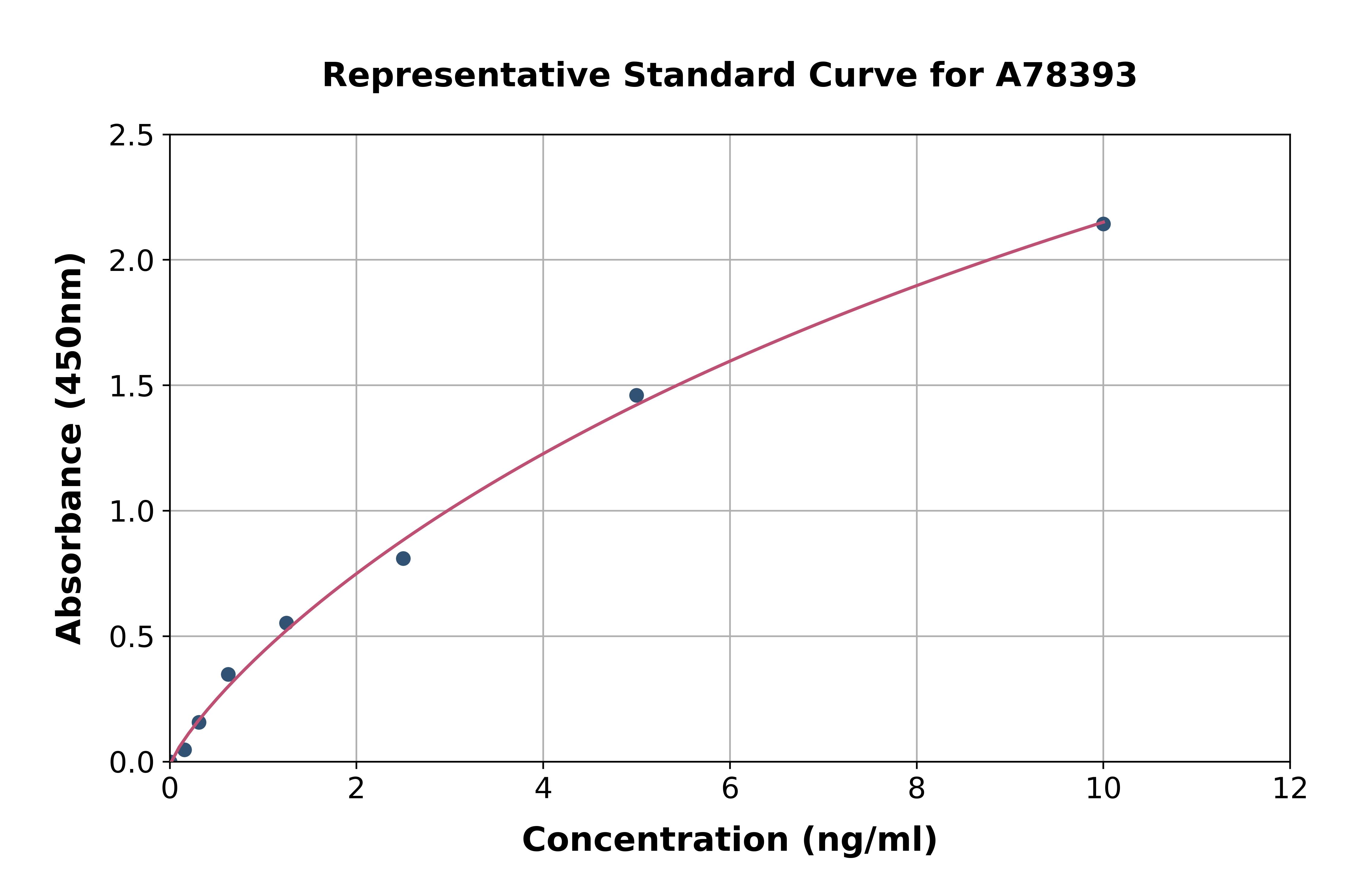
Rat LOX 1 ELISA KitA78393
Assay Sample TypePlasma, tissue homogenates and other biological fluids.
ReactivityRat
- SizePrice
Product group Assays
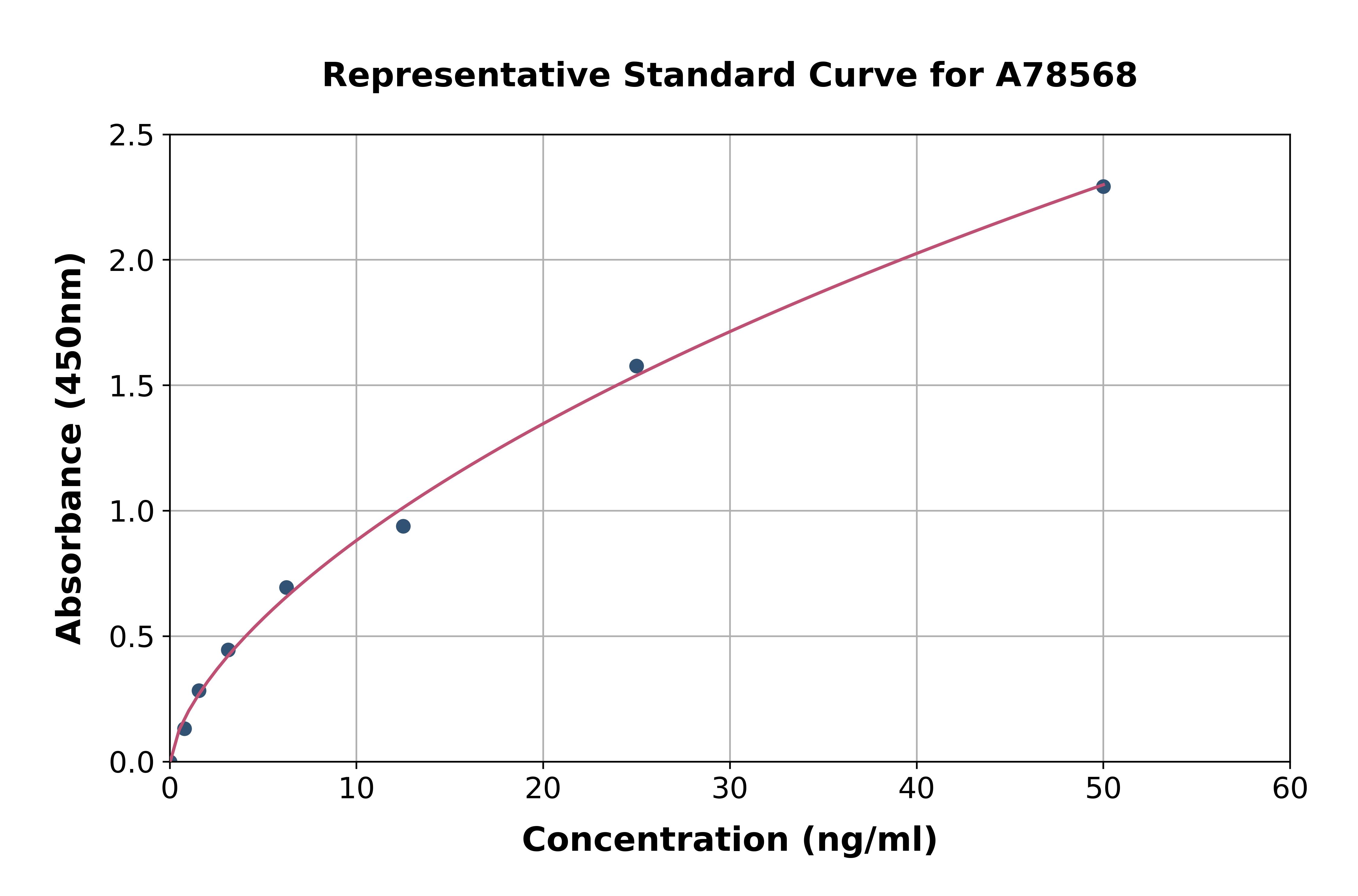
Mouse LOX 1 ELISA KitA78568
Assay Sample TypePlasma, tissue homogenates and other biological fluids.
ReactivityMouse
- SizePrice
Product group Assays
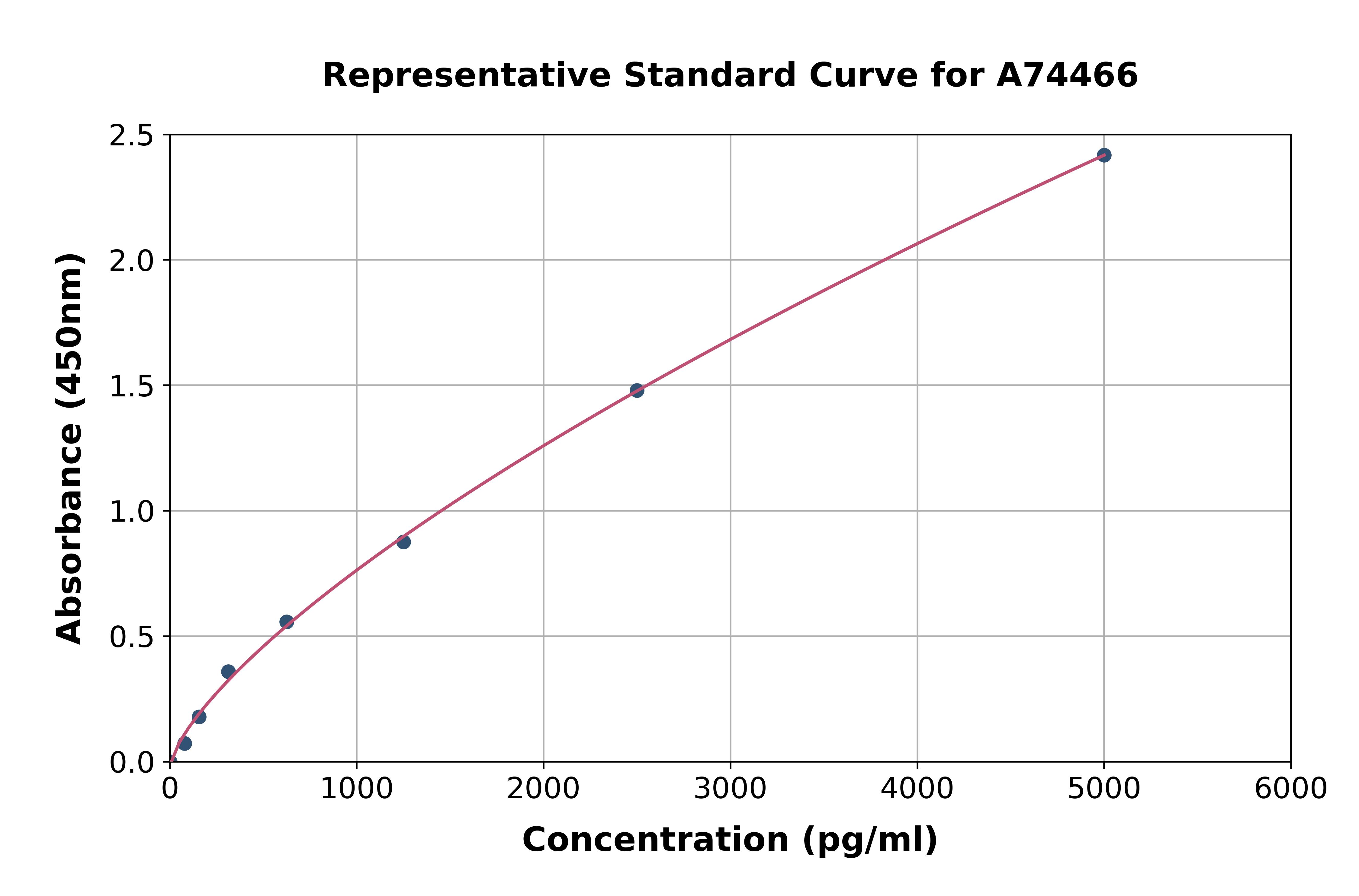
Rabbit LOX 1 ELISA KitA74466
Assay Sample TypePlasma, tissue homogenates and other biological fluids.
ReactivityRabbit
- SizePrice
Product group Assays
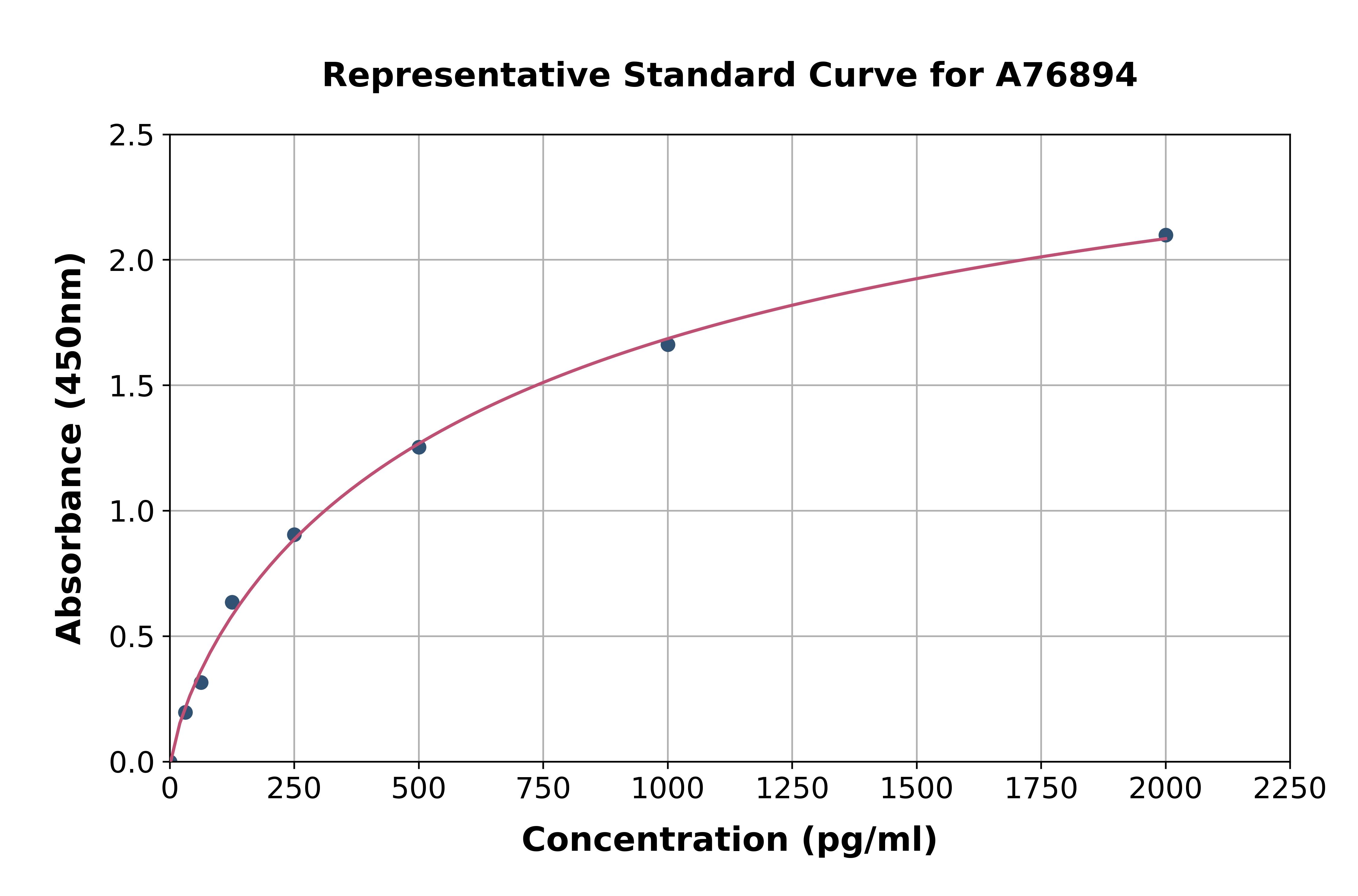
Human LOX 1 ELISA KitA76894
Assay Sample TypePlasma, tissue homogenates and other biological fluids.
ReactivityHuman
- SizePrice
Product group Antibodies
Anti-LOX (Center) Antibody102-10383
ApplicationsWestern Blot
TargetLOX
- SizePrice
Product group Antibodies
Anti-LOX (Center) Antibody102-27664
ApplicationsFlow Cytometry, Western Blot
TargetLOX
- SizePrice
Product group Assays
Assay Sample TypeSerum, plasma, tissue homogenates, cell lysates, cell culture supernates and other biological fluids.
ReactivityHuman
- SizePrice

Didn't find what you were looking for?
Search through our product groups to find the right product
Back to overview