

Search results: MAP2K1 MEK1
Product group DNA / RNA / Vectors
CategorycDNA
- SizePrice
Product group DNA / RNA / Vectors
CategorycDNA
- SizePrice
Product group DNA / RNA / Vectors
CategorycDNA
- SizePrice
Product group DNA / RNA / Vectors
CategorycDNA
- SizePrice
Product group DNA / RNA / Vectors
CategorycDNA
- SizePrice
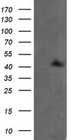
Product group Antibodies
ApplicationsImmunoFluorescence, Western Blot, ImmunoHistoChemistry
ReactivityCanine, Human, Monkey, Mouse, Rat
TargetMAP2K1
- SizePrice
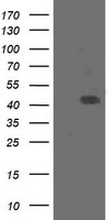
Product group Antibodies
ApplicationsImmunoFluorescence, Western Blot, ImmunoHistoChemistry
ReactivityCanine, Human, Monkey, Mouse, Rat
TargetMAP2K1
- SizePrice
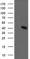
Product group Antibodies
ApplicationsImmunoFluorescence, Western Blot, ImmunoHistoChemistry
ReactivityCanine, Human, Monkey, Mouse, Rat
TargetMAP2K1
- SizePrice
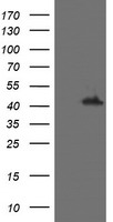
Product group Antibodies
ApplicationsImmunoFluorescence, Western Blot, ImmunoHistoChemistry
ReactivityCanine, Human, Monkey, Mouse, Rat
TargetMAP2K1
- SizePrice
Product group Antibodies
ApplicationsImmunoFluorescence, Western Blot, ImmunoHistoChemistry
ReactivityCanine, Human, Monkey, Mouse, Rat
TargetMAP2K1
- SizePrice
Product group Antibodies
ApplicationsImmunoFluorescence, Western Blot, ImmunoHistoChemistry
ReactivityCanine, Human, Monkey, Mouse, Rat
TargetMAP2K1
- SizePrice
Product group Antibodies
ApplicationsImmunoFluorescence, Western Blot, ImmunoHistoChemistry
ReactivityCanine, Human, Monkey, Mouse, Rat
TargetMAP2K1
- SizePrice
Didn't find what you were looking for?
Search through our product groups to find the right product
Back to overview