

Search results: MEKK2

Product group Antibodies
Anti-MEKK2/MAP3K2 Antibody Picoband(r)A02354-1-DYLIGHT488
ApplicationsFlow Cytometry, ImmunoFluorescence, Western Blot, ImmunoCytoChemistry
ReactivityHuman, Rat
TargetMAP3K2
- SizePrice
Product group Antibodies
Anti-MEKK2/MAP3K2 Antibody Picoband(r)A02354-1-DYLIGHT550
ApplicationsFlow Cytometry, ImmunoFluorescence, Western Blot, ImmunoCytoChemistry
ReactivityHuman, Rat
TargetMAP3K2
- SizePrice
Product group Antibodies
Anti-MEKK2/MAP3K2 Antibody Picoband(r)A02354-1-DYLIGHT594
ApplicationsFlow Cytometry, ImmunoFluorescence, Western Blot, ImmunoCytoChemistry
ReactivityHuman, Rat
TargetMAP3K2
- SizePrice
Product group Antibodies
Anti-MEKK2/MAP3K2 Antibody Picoband(r)A02354-1-FITC
ApplicationsFlow Cytometry, ImmunoFluorescence, Western Blot, ImmunoCytoChemistry
ReactivityHuman, Rat
TargetMAP3K2
- SizePrice
Product group Antibodies
Anti-MEKK2/MAP3K2 Antibody Picoband(r)A02354-1-HRP
ApplicationsFlow Cytometry, ImmunoFluorescence, Western Blot, ImmunoCytoChemistry
ReactivityHuman, Rat
TargetMAP3K2
- SizePrice
Product group Antibodies
Anti-MEKK2/MAP3K2 Antibody Picoband(r)A02354-1-IFLUOR647
ApplicationsFlow Cytometry, ImmunoFluorescence, Western Blot, ImmunoCytoChemistry
ReactivityHuman, Rat
TargetMAP3K2
- SizePrice
Product group Antibodies
Anti-MEKK2/MAP3K2 Antibody Picoband(r)A02354-1-PE
ApplicationsFlow Cytometry, ImmunoFluorescence, Western Blot, ImmunoCytoChemistry
ReactivityHuman, Rat
TargetMAP3K2
- SizePrice
Product group Antibodies
ApplicationsFlow Cytometry, ImmunoFluorescence, Western Blot, ImmunoCytoChemistry
ReactivityHuman, Rat
TargetMAP3K2
- SizePrice
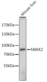
Product group Antibodies
ApplicationsImmunoPrecipitation, Western Blot
ReactivityHuman, Mouse
TargetMAP3K2
- SizePrice
Product group Antibodies
ApplicationsImmunoPrecipitation, Western Blot
ReactivityHuman
TargetMAP3K2
- SizePrice
Product group Antibodies
MEKK2 AntibodyCSB-PA942372XA01DOA
ApplicationsWestern Blot, ELISA
ReactivityPlant
- SizePrice
Didn't find what you were looking for?
Search through our product groups to find the right product
Back to overview