

Search results: MSL2
Product group Antibodies
ApplicationsImmunoPrecipitation
ReactivityHuman
- SizePrice
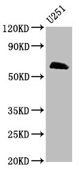
Product group Antibodies
ApplicationsWestern Blot
ReactivityHuman
- SizePrice
Product group Antibodies
MSL2 AntibodyCSB-PA704101XA01DOA
ApplicationsWestern Blot, ELISA
ReactivityPlant
TargetMSL2
- SizePrice
Product group Antibodies
MSL2 AntibodyCSB-PA875722LA01HU
ApplicationsWestern Blot, ELISA
ReactivityHuman
TargetMSL2
- SizePrice
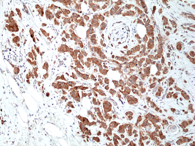
Product group Antibodies
anti-Musashi 2(MSl2) Rabbit Monoclonal (RM422)REV-31-1309-00
ApplicationsWestern Blot, ImmunoHistoChemistry
ReactivityHuman
TargetMSI2
- SizePrice

Product group Antibodies
Anti-MSL2 AntibodyHPA003413
ApplicationsWestern Blot
ReactivityHuman
TargetMSL2
- SizePrice
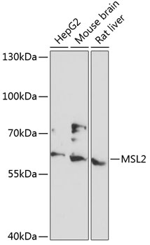
Product group Antibodies
Anti-MSL2 AntibodyA90763
ApplicationsWestern Blot
ReactivityHuman, Mouse, Rat
- SizePrice
Product group Antibodies
MSL2 Polyclonal AntibodyCAC15668
ApplicationsWestern Blot, ELISA
TargetMSL2
- SizePrice

Product group Antibodies
Anti-MSL2 AntibodyHPA048482
ApplicationsWestern Blot, ImmunoCytoChemistry
ReactivityHuman
TargetMSL2
- SizePrice
Didn't find what you were looking for?
Search through our product groups to find the right product
Back to overview