

Search results: PDCD4
Product group Antibodies
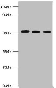
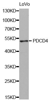
PDCD4 AntibodyCSB-PA703610DSR1HU
ApplicationsImmunoPrecipitation, Western Blot, ELISA
ReactivityHuman
TargetPDCD4
- SizePrice
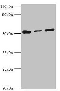
Product group Antibodies
PDCD4 AntibodyCSB-PA703610DSR2HU
ApplicationsImmunoPrecipitation, Western Blot, ELISA, ImmunoHistoChemistry
ReactivityHuman
TargetPDCD4
- SizePrice
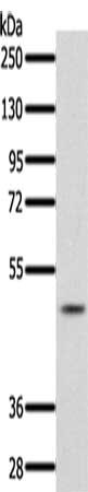
Product group Antibodies
PDCD4 AntibodyCSB-PA442649
ApplicationsWestern Blot, ELISA
ReactivityHuman, Mouse, Rat
TargetPDCD4
- SizePrice
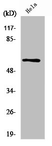
Product group Antibodies
PDCD4 AntibodyCSB-PA017670GA01HU
ApplicationsWestern Blot, ELISA, ImmunoHistoChemistry
ReactivityHuman, Mouse, Rat
TargetPDCD4
- SizePrice
Product group Antibodies
PDCD4 AntibodyCSB-PA003720
ApplicationsImmunoFluorescence, Western Blot, ELISA, ImmunoHistoChemistry
ReactivityHuman, Mouse, Rat
TargetPDCD4
- SizePrice
Product group Antibodies
PDCD4 AntibodyCSB-PA008871
ApplicationsELISA, ImmunoHistoChemistry
ReactivityHuman, Mouse, Rat
TargetPDCD4
- SizePrice

Product group Antibodies
Anti-PDCD4 AntibodyHPA001032
ApplicationsWestern Blot, ChIP Chromatin ImmunoPrecipitation, ImmunoHistoChemistry
ReactivityHuman, Mouse, Rat
TargetPDCD4
- SizePrice

Product group Antibodies
Anti-PDCD4 AntibodyA43332
ApplicationsWestern Blot
ReactivityHuman, Mouse, Rat
- SizePrice

Product group Antibodies
Anti-PDCD4 AntibodyA30394
ApplicationsWestern Blot, ImmunoHistoChemistry
ReactivityHuman, Mouse, Rat
- SizePrice
Didn't find what you were looking for?
Search through our product groups to find the right product
Back to overview