

Search results: PDCD4
Product group Assays
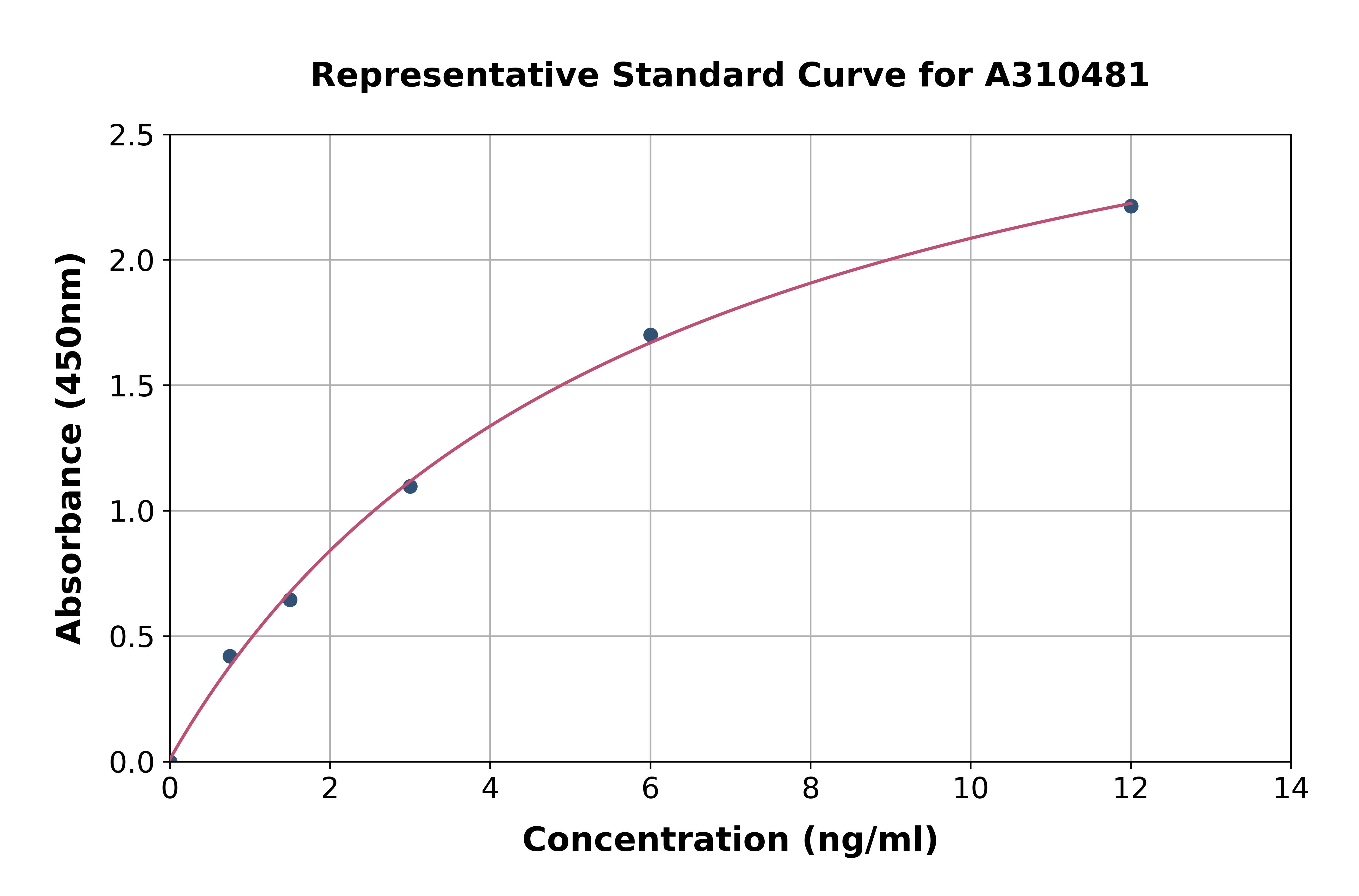
Pdcd4 ELISA KitEKA50464
Assay Sample TypeResearchers plate their cell line of choice
ReactivityHuman, Mouse, Rat
- SizePrice
Product group Antibodies
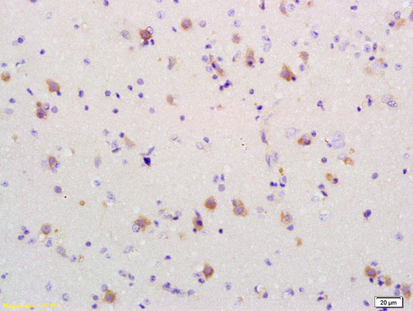
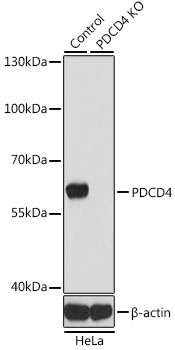
PDCD4 Polyclonal AntibodyBS-1608R
ApplicationsImmunoFluorescence, Western Blot, ELISA, ImmunoCytoChemistry, ImmunoHistoChemistry, ImmunoHistoChemistry Frozen, ImmunoHistoChemistry Paraffin
ReactivityHuman, Mouse, Rat
TargetPDCD4
- SizePrice
Product group Antibodies
PDCD4 Polyclonal AntibodyBS-55166R
ApplicationsImmunoFluorescence, Western Blot, ImmunoHistoChemistry, ImmunoHistoChemistry Frozen, ImmunoHistoChemistry Paraffin
ReactivityHuman, Mouse
TargetPDCD4
- SizePrice
Product group Antibodies
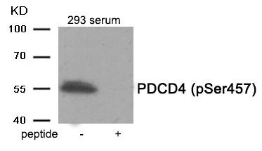
Phospho-PDCD4 (Ser457) AntibodyCSB-PA785011
ApplicationsWestern Blot, ELISA
ReactivityHuman, Mouse, Rat
TargetPDCD4
- SizePrice
![Anti-PDCD4 Antibody [ARC1398]](https://www.antibodies.com/image/catalog/307/A307976_1.jpg)
Product group Antibodies
ApplicationsWestern Blot, ImmunoHistoChemistry
ReactivityHuman, Mouse, Rat
- SizePrice
Product group Antibodies
Phospho-PDCD4 (S67) AntibodyCSB-PA008869
ApplicationsWestern Blot, ELISA, ImmunoHistoChemistry
ReactivityHuman, Monkey, Mouse, Rat
TargetPDCD4
- SizePrice
Product group Antibodies
Phospho-PDCD4 (S457) AntibodyCSB-PA008870
ApplicationsWestern Blot, ELISA, ImmunoHistoChemistry
ReactivityHuman, Mouse, Rat
TargetPDCD4
- SizePrice
Product group Antibodies
ApplicationsWestern Blot, ELISA
ReactivityHuman
- SizePrice
Product group Proteins / Signaling Molecules
ApplicationsOther Application
- SizePrice
Product group Antibodies
ApplicationsImmunoFluorescence, Western Blot, ImmunoHistoChemistry
ReactivityHuman, Mouse, Rat
- SizePrice
Didn't find what you were looking for?
Search through our product groups to find the right product
Back to overview