

Search results: RALB
![IHC-P analysis of breast adenocarcinoma tissue using GTX83734 RALBP1 antibody [2A6]. Antigen retrieval : Heat-induced epitope retrieval by 10mM citrate buffer, pH6.0, 100oC for 10min. Dilution : 1:50](https://www.genetex.com/upload/website/prouct_img/normal/GTX83734/GTX83734_1771_IHC-P_w_23061420_538.webp)
Product group Antibodies
RALBP1 antibody [2A6]GTX83734
ApplicationsImmunoFluorescence, ImmunoPrecipitation, Western Blot, ImmunoCytoChemistry, ImmunoHistoChemistry, ImmunoHistoChemistry Paraffin
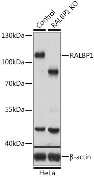
TargetRALBP1
- SizePrice
![ICC/IF analysis of U2OS cells using GTX03249 RALBP1 antibody [GT1337]. Blue : DAPI for nuclear staining Dilution : 1:100](https://www.genetex.com/upload/website/prouct_img/normal/GTX03249/GTX03249_20210615_ICCIF_81_w_23053123_675.webp)
Product group Antibodies
RALBP1 antibody [GT1337]GTX03249
ApplicationsImmunoFluorescence, Western Blot, ImmunoCytoChemistry, ImmunoHistoChemistry, ImmunoHistoChemistry Paraffin
TargetRALBP1
- SizePrice
Product group Assays
Assay Sample TypeTissue Homogenates, Cell Lysates And Other Biological Fluids
- SizePrice
Product group Assays
Assay Sample TypeTissue Homogenates, Cell Lysates And Other Biological Fluids
- SizePrice
Product group Antibodies
RalBP1 antibodyGTX55774
ApplicationsWestern Blot, ImmunoHistoChemistry, ImmunoHistoChemistry Paraffin
TargetRALBP1
- SizePrice
Product group Assays
Assay Sample TypeSerum, Plasma
- SizePrice
![FACS analysis of HeLa cells using GTX83726 RALBP1 antibody [6C6]. Red : Primary antibody Blue : Negative control antibody](https://www.genetex.com/upload/website/prouct_img/normal/GTX83726/GTX83726_154_FACS_w_23061420_353.webp)
Product group Antibodies
RALBP1 antibody [6C6]GTX83726
ApplicationsFlow Cytometry, ImmunoFluorescence, ImmunoPrecipitation, Western Blot, ImmunoCytoChemistry
TargetRALBP1
- SizePrice
Didn't find what you were looking for?
Search through our product groups to find the right product
Back to overview