

Search results: RALB
![FACS analysis of HEK293T cells transfected with either RALBP1 plasmid(Red) or empty vector control plasmid(Blue) using GTX83727 RALBP1 antibody [1H9].](https://www.genetex.com/upload/website/prouct_img/normal/GTX83727/GTX83727_156_FACS_w_23061420_352.webp)
Product group Antibodies
RALBP1 antibody [1H9]GTX83727
ApplicationsFlow Cytometry, ImmunoPrecipitation, Western Blot
TargetRALBP1
- SizePrice
![FACS analysis of Jurkat cells using GTX83728 RALBP1 antibody [11B2]. Red : Primary antibody Blue : Negative control antibody](https://www.genetex.com/upload/website/prouct_img/normal/GTX83728/GTX83728_157_FACS_w_23061420_844.webp)
Product group Antibodies
RALBP1 antibody [11B2]GTX83728
ApplicationsFlow Cytometry, ImmunoFluorescence, ImmunoPrecipitation, Western Blot, ImmunoCytoChemistry
TargetRALBP1
- SizePrice
![FACS analysis of HEK293T cells transfected with either RALBP1 plasmid(Red) or empty vector control plasmid(Blue) using GTX83730 RALBP1 antibody [6G10].](https://www.genetex.com/upload/website/prouct_img/normal/GTX83730/GTX83730_159_FACS_w_23061420_496.webp)
Product group Antibodies
RALBP1 antibody [6G10]GTX83730
ApplicationsFlow Cytometry, ImmunoFluorescence, ImmunoPrecipitation, Western Blot, ImmunoCytoChemistry, ImmunoHistoChemistry, ImmunoHistoChemistry Paraffin
TargetRALBP1
- SizePrice
Product group Antibodies
RALBP1 AntibodyCSB-PA019298GA01HU
ApplicationsWestern Blot, ELISA
TargetRALBP1
- SizePrice
Product group Antibodies
RALBP1 AntibodyCSB-PA618981LA01HU
ApplicationsImmunoFluorescence, Western Blot, ELISA, ImmunoHistoChemistry
TargetRALBP1
- SizePrice

Product group Antibodies
Anti-RALBP1 AntibodyHPA046651
ApplicationsImmunoCytoChemistry, ImmunoHistoChemistry
TargetRALBP1
- SizePrice

Product group Antibodies
Anti-RALBP1 AntibodyHPA007622
ApplicationsImmunoCytoChemistry
TargetRALBP1
- SizePrice
Product group Antibodies

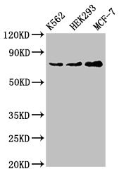
ApplicationsWestern Blot
- SizePrice
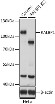
Product group Antibodies
ApplicationsWestern Blot, ELISA, ImmunoHistoChemistry
- SizePrice
Product group DNA / RNA / Vectors
Human, RALBP1 cDNA ORF Clone, untaggedHG11178-UT
CategoryDNA / RNA / Vectors
- SizePrice
Product group Antibodies
RALBP1 Polyclonal AntibodyBS-55186R
ApplicationsWestern Blot, ImmunoHistoChemistry, ImmunoHistoChemistry Paraffin
TargetRALBP1
- SizePrice
Didn't find what you were looking for?
Search through our product groups to find the right product
Back to overview