

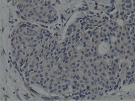
IHC-P analysis of human breast adenocarcinoma tissue using GTX83658 SH3GL1 antibody [3F8]. Antigen retrieval : Heat-induced epitope retrieval by 10mM citrate buffer, pH6.0, 100oC for 10min.
![WB analysis of HEK293T cells transfected with SH3GL1 plasmid (Right) or empty vector (Left) for 48 hrs using GTX83658 SH3GL1 antibody [3F8]. Loading : 5 ug per lane](https://www.genetex.com/upload/website/prouct_img/normal/GTX83658/GTX83658_3799_WB_w_23061420_987.webp "WB analysis of HEK293T cells transfected with SH3GL1 plasmid (Right) or empty vector (Left) for 48 hrs using GTX83658 SH3GL1 antibody [3F8]. Loading : 5 ug per lane")
![WB analysis of various cell lines using GTX83658 SH3GL1 antibody [3F8]. Loading : 35 ug per lane Dilution : 1:200](https://www.genetex.com/upload/website/prouct_img/normal/GTX83658/GTX83658_3442_WB_w_23061420_334.webp "WB analysis of various cell lines using GTX83658 SH3GL1 antibody [3F8]. Loading : 35 ug per lane Dilution : 1:200")
![ICC/IF analysis of COS7 cells transiently transfected with SH3GL1 plasmid using GTX83658 SH3GL1 antibody [3F8].](https://www.genetex.com/upload/website/prouct_img/normal/GTX83658/GTX83658_765_ICCIF_w_23061420_822.webp "ICC/IF analysis of COS7 cells transiently transfected with SH3GL1 plasmid using GTX83658 SH3GL1 antibody [3F8].")
![IHC-P analysis of human bladder carcinoma tissue using GTX83658 SH3GL1 antibody [3F8]. Antigen retrieval : Heat-induced epitope retrieval by 10mM citrate buffer, pH6.0, 100oC for 10min.](https://www.genetex.com/upload/website/prouct_img/normal/GTX83658/GTX83658_1676_IHC-P_w_23061420_101.webp "IHC-P analysis of human bladder carcinoma tissue using GTX83658 SH3GL1 antibody [3F8]. Antigen retrieval : Heat-induced epitope retrieval by 10mM citrate buffer, pH6.0, 100oC for 10min.")
![WB analysis of wild type and SH3GL1 knock out 293T cell lysate(10μg per lane) using GTX83658 SH3GL1 antibody [3F8]. Dilution : 1:500](https://www.genetex.com/upload/website/prouct_img/normal/GTX83658/GTX83658_3798_20181228_WB_w_23061420_374.webp "WB analysis of wild type and SH3GL1 knock out 293T cell lysate(10μg per lane) using GTX83658 SH3GL1 antibody [3F8]. Dilution : 1:500")
IHC-P analysis of human breast adenocarcinoma tissue using GTX83658 SH3GL1 antibody [3F8]. Antigen retrieval : Heat-induced epitope retrieval by 10mM citrate buffer, pH6.0, 100oC for 10min.
SH3GL1 antibody [3F8]
GTX83658
ApplicationsImmunoFluorescence, Western Blot, ImmunoCytoChemistry, ImmunoHistoChemistry, ImmunoHistoChemistry Paraffin
Product group Antibodies
ReactivityHuman, Monkey, Mouse
TargetSH3GL1
Overview
- SupplierGeneTex
- Product NameSH3GL1 antibody [3F8]
- Delivery Days Customer9
- Application Supplier NoteWB: 1:2000. ICC/IF: 1:100. IHC-P: 1:150. *Optimal dilutions/concentrations should be determined by the researcher.Not tested in other applications.
- ApplicationsImmunoFluorescence, Western Blot, ImmunoCytoChemistry, ImmunoHistoChemistry, ImmunoHistoChemistry Paraffin
- CertificationResearch Use Only
- ClonalityMonoclonal
- Clone ID3F8
- Concentration0.65 mg/ml
- ConjugateUnconjugated
- Gene ID6455
- Target nameSH3GL1
- Target descriptionSH3 domain containing GRB2 like 1, endophilin A2
- Target synonymsCNSA1, EEN, SH3D2B, SH3P8, endophilin-A2, EEN fusion partner of MLL, SH3 domain protein 2B, SH3 domain-containing GRB2-like protein 1, SH3-containing Grb-2-like 1 protein, SH3-domain GRB2-like 1, endophilin-2, extra 11-19 leukemia fusion, extra eleven-nineteen leukemia fusion gene protein
- HostMouse
- IsotypeIgG1
- Protein IDQ99961
- Protein NameEndophilin-A2
- Scientific DescriptionImplicated in endocytosis. May recruit other proteins to membranes with high curvature.
- ReactivityHuman, Monkey, Mouse
- Storage Instruction-20°C or -80°C,2°C to 8°C
- UNSPSC41116161
Datasheet
Related products

Product group Antibodies
ApplicationsWestern Blot, ImmunoHistoChemistry
ReactivityHuman, Monkey, Mouse, Rat
TargetSH3GL1
- SizePrice
Product group Antibodies
Anti-SH3GL1 AntibodyA32074
ApplicationsWestern Blot, ImmunoHistoChemistry
ReactivityHuman, Mouse, Rat
- SizePrice
Product group Antibodies

Goat anti-Endophilin-A2EB10691
ApplicationsWestern Blot, ELISA, ImmunoHistoChemistry
ReactivityBovine, Canine, Human
TargetSH3GL1
- SizePrice

Product group Antibodies
Anti-SH3GL1 AntibodyHPA021485
ApplicationsWestern Blot, ImmunoCytoChemistry, ImmunoHistoChemistry
ReactivityHuman
TargetSH3GL1
- SizePrice
Product group Antibodies

SH3GL1 / EEN AntibodyLS-C349151
ApplicationsWestern Blot, ImmunoHistoChemistry
ReactivityHuman, Monkey, Mouse, Rat
TargetSH3GL1
- SizePrice
Product group Antibodies
SH3GL1 AntibodyCSB-PA859949ESR1HU
ApplicationsWestern Blot, ELISA, ImmunoHistoChemistry
ReactivityHuman
TargetSH3GL1
- SizePrice
Product group Antibodies
SH3GL1 antibodyGTX66494
ApplicationsWestern Blot
ReactivityHuman, Monkey
TargetSH3GL1
- SizePrice
![Various whole cell extracts (30 μg) were separated by 10% SDS-PAGE, and the membrane was blotted with SH3GL1 antibody [2F5] (GTX83657) diluted at 1:1000. The HRP-conjugated anti-mouse IgG antibody (GTX213111-01) was used to detect the primary antibody, and the signal was developed with Trident ECL plus-Enhanced. Corresponding RNA expression data for the same cell lines are based on Human Protein Atlas program.](https://www.genetex.com/upload/website/prouct_img/normal/GTX83657/GTX83657_822205142_20221202_WB_TPM_watermark_22120719_701.webp)
Product group Antibodies
SH3GL1 antibody [2F5]GTX83657
ApplicationsFlow Cytometry, ImmunoFluorescence, Western Blot, ImmunoCytoChemistry, ImmunoHistoChemistry, ImmunoHistoChemistry Paraffin
ReactivityHuman
TargetSH3GL1
- SizePrice