

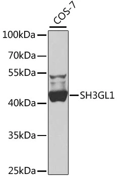
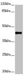
WB analysis of COS-7 cell lysate using GTX66494 SH3GL1 antibody. Dilution : 1:1000 Loading : 25μg per lane
WB analysis of COS-7 cell lysate using GTX66494 SH3GL1 antibody. Dilution : 1:1000 Loading : 25μg per lane
SH3GL1 antibody
GTX66494
ApplicationsWestern Blot
Product group Antibodies
ReactivityHuman, Monkey
TargetSH3GL1
Overview
- SupplierGeneTex
- Product NameSH3GL1 antibody
- Delivery Days Customer9
- Application Supplier NoteWB: 1:500 - 1:2000. *Optimal dilutions/concentrations should be determined by the researcher.Not tested in other applications.
- ApplicationsWestern Blot
- CertificationResearch Use Only
- ClonalityPolyclonal
- ConjugateUnconjugated
- Gene ID6455
- Target nameSH3GL1
- Target descriptionSH3 domain containing GRB2 like 1, endophilin A2
- Target synonymsCNSA1, EEN, SH3D2B, SH3P8, endophilin-A2, EEN fusion partner of MLL, SH3 domain protein 2B, SH3 domain-containing GRB2-like protein 1, SH3-containing Grb-2-like 1 protein, SH3-domain GRB2-like 1, endophilin-2, extra 11-19 leukemia fusion, extra eleven-nineteen leukemia fusion gene protein
- HostRabbit
- IsotypeIgG
- Protein IDQ99961
- Protein NameEndophilin-A2
- Scientific DescriptionThis gene encodes a member of the endophilin family of Src homology 3 domain-containing proteins. The encoded protein is involved in endocytosis and may also play a role in the cell cycle. Overexpression of this gene may play a role in leukemogenesis, and the encoded protein has been implicated in acute myeloid leukemia as a fusion partner of the myeloid-lymphoid leukemia protein. Pseudogenes of this gene are located on the long arm of chromosomes 11 and 17. Alternatively spliced transcript variants encoding multiple isoforms have been observed for this gene. [provided by RefSeq, Jan 2011]
- ReactivityHuman, Monkey
- Storage Instruction-20°C or -80°C,2°C to 8°C
- UNSPSC41116161
Datasheet
Related products

Product group Antibodies
ApplicationsWestern Blot, ImmunoHistoChemistry
ReactivityHuman, Monkey, Mouse, Rat
TargetSH3GL1
- SizePrice
Product group Antibodies
Anti-SH3GL1 AntibodyA32074
ApplicationsWestern Blot, ImmunoHistoChemistry
ReactivityHuman, Mouse, Rat
- SizePrice
Product group Antibodies

Goat anti-Endophilin-A2EB10691
ApplicationsWestern Blot, ELISA, ImmunoHistoChemistry
ReactivityBovine, Canine, Human
TargetSH3GL1
- SizePrice

Product group Antibodies
Anti-SH3GL1 AntibodyHPA021485
ApplicationsWestern Blot, ImmunoCytoChemistry, ImmunoHistoChemistry
ReactivityHuman
TargetSH3GL1
- SizePrice
Product group Antibodies

SH3GL1 / EEN AntibodyLS-C349151
ApplicationsWestern Blot, ImmunoHistoChemistry
ReactivityHuman, Monkey, Mouse, Rat
TargetSH3GL1
- SizePrice
Product group Antibodies
SH3GL1 AntibodyCSB-PA859949ESR1HU
ApplicationsWestern Blot, ELISA, ImmunoHistoChemistry
ReactivityHuman
TargetSH3GL1
- SizePrice
![Various whole cell extracts (30 μg) were separated by 10% SDS-PAGE, and the membrane was blotted with SH3GL1 antibody [2F5] (GTX83657) diluted at 1:1000. The HRP-conjugated anti-mouse IgG antibody (GTX213111-01) was used to detect the primary antibody, and the signal was developed with Trident ECL plus-Enhanced. Corresponding RNA expression data for the same cell lines are based on Human Protein Atlas program.](https://www.genetex.com/upload/website/prouct_img/normal/GTX83657/GTX83657_822205142_20221202_WB_TPM_watermark_22120719_701.webp)
Product group Antibodies
SH3GL1 antibody [2F5]GTX83657
ApplicationsFlow Cytometry, ImmunoFluorescence, Western Blot, ImmunoCytoChemistry, ImmunoHistoChemistry, ImmunoHistoChemistry Paraffin
ReactivityHuman
TargetSH3GL1
- SizePrice
![IHC-P analysis of human breast adenocarcinoma tissue using GTX83658 SH3GL1 antibody [3F8]. Antigen retrieval : Heat-induced epitope retrieval by 10mM citrate buffer, pH6.0, 100oC for 10min.](https://www.genetex.com/upload/website/prouct_img/normal/GTX83658/GTX83658_1675_IHC-P_w_23061420_442.webp)
Product group Antibodies
SH3GL1 antibody [3F8]GTX83658
ApplicationsImmunoFluorescence, Western Blot, ImmunoCytoChemistry, ImmunoHistoChemistry, ImmunoHistoChemistry Paraffin
ReactivityHuman, Monkey, Mouse
TargetSH3GL1
- SizePrice