

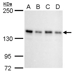
SIN3A antibody detects SIN3A protein by western blot analysis. A 293T whole cell lysate/extract B A431 whole cell lysate/extract C HeLa whole cell lysate/extract D HepG2 whole cell lysate/extract 5% SDS-PAGE SIN3A antibody (GTX129156) dilution: 1:1000 The HRP-conjugated anti-rabbit IgG antibody (GTX213110-01) was used to detect the primary antibody.
 and 5 μg of either control rabbit IgG or anti-SIN3A antibody. The precipitated DNA was detected by PCR with primer set targeting to GAPDH or MyoD.")
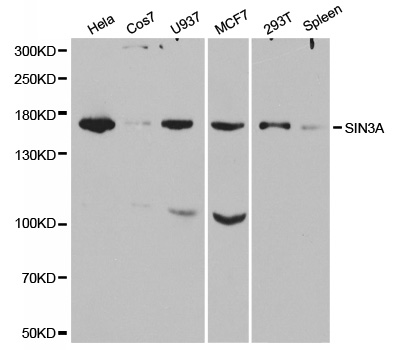
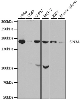
 were separated by 5% SDS-PAGE, and the membrane was blotted with SIN3A antibody (GTX129156) diluted at 1:1000. The HRP-conjugated anti-rabbit IgG antibody (GTX213110-01) was used to detect the primary antibody.")
 diluted at 1:1000. Red: phalloidin, a cytoskeleton marker, diluted at 1:200. Scale bar= 10 μm.")
 dilution: 1:500.
Antigen Retrieval: Trilogy? (EDTA based, pH 8.0) buffer, 15min")
 were separated by 5% SDS-PAGE, and the membrane was blotted with SIN3A antibody (GTX129156) diluted at 1:1000. The HRP-conjugated anti-rabbit IgG antibody (GTX213110-01) was used to detect the primary antibody.")
 dilution: 1:500.
Antigen Retrieval: Trilogy? (EDTA based, pH 8.0) buffer, 15min")
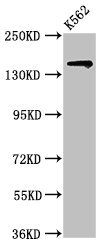
 5% SDS-PAGE The immunoprecipitated SIN3A protein was detected by SIN3A antibody (GTX129156) diluted at 1 : 1000. EasyBlot anti-rabbit IgG (HRP) (GTX221666-01) was used as a secondary reagent.")
 dilution: 1:500.
Antigen Retrieval: Trilogy? (EDTA based, pH 8.0) buffer, 15min")
SIN3A antibody detects SIN3A protein by western blot analysis. A 293T whole cell lysate/extract B A431 whole cell lysate/extract C HeLa whole cell lysate/extract D HepG2 whole cell lysate/extract 5% SDS-PAGE SIN3A antibody (GTX129156) dilution: 1:1000 The HRP-conjugated anti-rabbit IgG antibody (GTX213110-01) was used to detect the primary antibody.
SIN3A antibody
GTX129156
ApplicationsImmunoFluorescence, ImmunoPrecipitation, Western Blot, ChIP Chromatin ImmunoPrecipitation, ImmunoCytoChemistry, ImmunoHistoChemistry, ImmunoHistoChemistry Paraffin
Product group Antibodies
ReactivityHuman, Mouse, Rat
TargetSIN3A
Overview
- SupplierGeneTex
- Product NameSIN3A antibody
- Delivery Days Customer9
- Application Supplier NoteWB: 1:500-1:3000. ICC/IF: 1:100-1:1000. IHC-P: 1:100-1:1000. IP: 1:100-1:500. *Optimal dilutions/concentrations should be determined by the researcher.Not tested in other applications.
- ApplicationsImmunoFluorescence, ImmunoPrecipitation, Western Blot, ChIP Chromatin ImmunoPrecipitation, ImmunoCytoChemistry, ImmunoHistoChemistry, ImmunoHistoChemistry Paraffin
- CertificationResearch Use Only
- ClonalityPolyclonal
- Concentration1.38 mg/ml
- ConjugateUnconjugated
- Gene ID25942
- Target nameSIN3A
- Target descriptionSIN3 transcription regulator family member A
- Target synonymsCHR15DELq24, DEL15Q24, WITKOS, paired amphipathic helix protein Sin3a, SIN3 homolog A, transcription regulator, histone deacetylase complex subunit Sin3a, transcriptional co-repressor Sin3A, transcriptional corepressor Sin3a, transcriptional regulator, SIN3A
- HostRabbit
- IsotypeIgG
- Protein IDQ96ST3
- Protein NamePaired amphipathic helix protein Sin3a
- Scientific DescriptionThe protein encoded by this gene is a transcriptional regulatory protein. It contains paired amphipathic helix (PAH) domains, which are important for protein-protein interactions and may mediate repression by the Mad-Max complex. [provided by RefSeq]
- ReactivityHuman, Mouse, Rat
- Storage Instruction-20°C or -80°C,2°C to 8°C
- UNSPSC41116161
Datasheet
Related products

Product group Antibodies
Anti-mSin3A/SIN3A Antibody Picoband(r)A01203-2-CARRIER-FREE
ApplicationsFlow Cytometry, ImmunoFluorescence, Western Blot, ELISA, ImmunoCytoChemistry, ImmunoHistoChemistry
ReactivityHuman, Rat
TargetSIN3A
- SizePrice
Product group Antibodies
Anti-SIN3A AntibodyA30485
ApplicationsImmunoFluorescence, ImmunoPrecipitation, Western Blot, ChIP Chromatin ImmunoPrecipitation, ImmunoHistoChemistry
ReactivityHuman, Mouse, Rat
- SizePrice
Product group Antibodies

SIN3A Antibody (aa1-120)LS-C748945
ApplicationsWestern Blot, ImmunoHistoChemistry
ReactivityHuman, Rat
TargetSIN3A
- SizePrice

Product group Antibodies
Anti-SIN3A AntibodyHPA047213
ApplicationsImmunoHistoChemistry
ReactivityHuman
TargetSIN3A
- SizePrice
Product group Antibodies
SIN3A AntibodyCSB-PA836303LA01HU
ApplicationsWestern Blot, ELISA, ImmunoHistoChemistry
ReactivityHuman
TargetSIN3A
- SizePrice
Product group Antibodies
SIN3A antibodyGTX23479
ApplicationsImmunoFluorescence, ImmunoPrecipitation, Western Blot, ChIP Chromatin ImmunoPrecipitation, ImmunoCytoChemistry, ImmunoHistoChemistry, ImmunoHistoChemistry Paraffin
ReactivityHuman, Mouse, Rat
TargetSIN3A
- SizePrice
Product group Antibodies
SIN3A Polyclonal AntibodyCAC15027
ApplicationsWestern Blot, ELISA, ImmunoHistoChemistry
TargetSIN3A
- SizePrice
Product group Antibodies
SIN3A antibodyGTX32874
ApplicationsImmunoFluorescence, ImmunoPrecipitation, Western Blot, ImmunoCytoChemistry
ReactivityHuman, Monkey, Mouse
TargetSIN3A
- SizePrice