


Western Blot analysis of Jurkat cells using Smad1 Polyclonal Antibody
Western Blot analysis of Jurkat cells using Smad1 Polyclonal Antibody
SMAD1 Antibody
CSB-PA004104
ApplicationsImmunoFluorescence, Western Blot, ELISA, ImmunoHistoChemistry
Product group Antibodies
ReactivityHuman, Mouse
TargetSMAD1
Overview
- SupplierCusabio
- Product NameSMAD1 Antibody
- Delivery Days Customer20
- ApplicationsImmunoFluorescence, Western Blot, ELISA, ImmunoHistoChemistry
- CertificationResearch Use Only
- ClonalityPolyclonal
- ConjugateUnconjugated
- Gene ID4086
- Target nameSMAD1
- Target descriptionSMAD family member 1
- Target synonymsBSP-1, BSP1, JV4-1, JV41, MADH1, MADR1, mothers against decapentaplegic homolog 1, MAD, mothers against decapentaplegic homolog 1, Mad-related protein 1, SMAD, mothers against DPP homolog 1, TGF-beta signaling protein 1, mothers against DPP homolog 1, transforming growth factor-beta signaling protein 1
- HostRabbit
- IsotypeIgG
- Protein IDQ15797
- Protein NameMothers against decapentaplegic homolog 1
- ReactivityHuman, Mouse
- Storage Instruction-20°C or -80°C
- UNSPSC41116161
Related products
Product group Antibodies
Anti-Smad1 AntibodyA95781
ApplicationsWestern Blot, ELISA, ImmunoHistoChemistry
ReactivityHuman, Mouse, Rat
- SizePrice

Product group Antibodies
Anti-SMAD1/SMAD5 Antibody Picoband(r)A00728-1-CARRIER-FREE
ApplicationsWestern Blot
ReactivityHuman, Mouse, Rat
TargetSMAD1
- SizePrice
Product group Antibodies
Anti-Smad1 Antibody130-00004
ApplicationsWestern Blot, ELISA
ReactivityHuman
TargetSMAD1
- SizePrice
Product group Antibodies
References
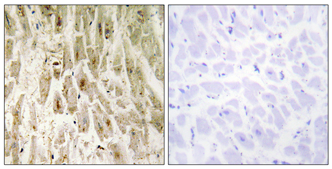
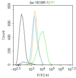
Smad1 Polyclonal AntibodyBS-1619R
ApplicationsFlow Cytometry, ImmunoFluorescence, ELISA, ImmunoCytoChemistry, ImmunoHistoChemistry, ImmunoHistoChemistry Frozen, ImmunoHistoChemistry Paraffin
ReactivityBovine, Canine, Equine, Human, Mouse, Porcine, Rabbit, Rat
TargetSMAD1
- SizePrice
Product group Antibodies
SMAD1 Polyclonal AntibodyCAC14179
ApplicationsImmunoFluorescence, Western Blot, ELISA, ImmunoHistoChemistry
TargetSMAD1
- SizePrice
Product group Antibodies

ApplicationsWestern Blot, ELISA
ReactivityHuman
TargetSMAD1
- SizePrice
Product group Antibodies
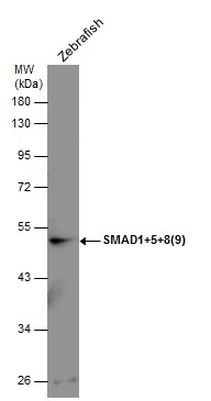
SMAD1+5+8(9) antibodyGTX131100
ApplicationsWestern Blot
ReactivityZebra Fish
- SizePrice
Product group Antibodies
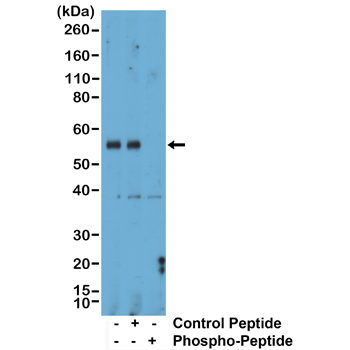
anti-Phospho-Smad1 (Ser463/465) / Smad5 (Ser463/465) / Smad9 (Ser465/467), Rabbit Monoclonal (RM487)REV-31-1379-00
ApplicationsWestern Blot
ReactivityHuman, Mouse, Rat
TargetSMAD1
- SizePrice