

WB (peptide competition) analysis of HepG2 cells stimulated with TGF beta using GTX25487 SMAD2 (phospho Thr8) antibody prior incubated with the non-phosphopeptide corresponding to the immunogen (Lane 2), a generic phosphothreonine containing peptide (Lane 3), or, the phosphopeptide immunogen (Lane 4) control. The data show that only the immunogen phosphopeptide blocks the signal, demonstrating the specificity of the antibody. The membrane treated with phosphatase (Lane 5) eliminates the signal further verifying that the antibody is phospho-specific.
 antibody. Normal Rabbit IgG was used as a negative IP control. The precipitated DNA was detected by PCR with primer set targeting to the promoter of ID1 gene as positive control and the inactive SAT2 used as negative control target.")
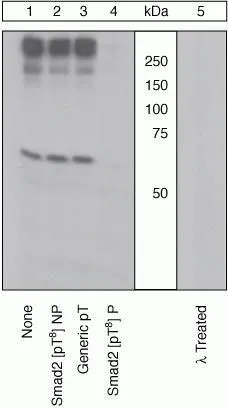
WB (peptide competition) analysis of HepG2 cells stimulated with TGF beta using GTX25487 SMAD2 (phospho Thr8) antibody prior incubated with the non-phosphopeptide corresponding to the immunogen (Lane 2), a generic phosphothreonine containing peptide (Lane 3), or, the phosphopeptide immunogen (Lane 4) control. The data show that only the immunogen phosphopeptide blocks the signal, demonstrating the specificity of the antibody. The membrane treated with phosphatase (Lane 5) eliminates the signal further verifying that the antibody is phospho-specific.
SMAD2 (phospho Thr8) antibody
GTX25487
ApplicationsFlow Cytometry, Western Blot, ChIP Chromatin ImmunoPrecipitation
Product group Antibodies
ReactivityHuman, Mouse, Rat
TargetSMAD2
Overview
- SupplierGeneTex
- Product NameSMAD2 (phospho Thr8) antibody
- Delivery Days Customer9
- Application Supplier NoteFCM: 1:20. ChIP assay: 10microl. *Optimal dilutions/concentrations should be determined by the researcher.Not tested in other applications.
- ApplicationsFlow Cytometry, Western Blot, ChIP Chromatin ImmunoPrecipitation
- CertificationResearch Use Only
- ClonalityPolyclonal
- ConjugateUnconjugated
- Gene ID4087
- Target nameSMAD2
- Target descriptionSMAD family member 2
- Target synonymsCHTD8, JV18, JV18-1, LDS6, MADH2, MADR2, hMAD-2, hSMAD2, mothers against decapentaplegic homolog 2, MAD homolog 2, SMAD, mothers against DPP homolog 2, Sma- and Mad-related protein 2, mother against DPP homolog 2
- HostRabbit
- IsotypeIgG
- Protein IDQ15796
- Protein NameMothers against decapentaplegic homolog 2
- Scientific DescriptionThe protein encoded by this gene belongs to the SMAD, a family of proteins similar to the gene products of the Drosophila gene mothers against decapentaplegic (Mad) and the C. elegans gene Sma. SMAD proteins are signal transducers and transcriptional modulators that mediate multiple signaling pathways. This protein mediates the signal of the transforming growth factor (TGF)-beta, and thus regulates multiple cellular processes, such as cell proliferation, apoptosis, and differentiation. This protein is recruited to the TGF-beta receptors through its interaction with the SMAD anchor for receptor activation (SARA) protein. In response to TGF-beta signal, this protein is phosphorylated by the TGF-beta receptors. The phosphorylation induces the dissociation of this protein with SARA and the association with the family member SMAD4. The association with SMAD4 is important for the translocation of this protein into the nucleus, where it binds to target promoters and forms a transcription repressor complex with other cofactors. This protein can also be phosphorylated by activin type 1 receptor kinase, and mediates the signal from the activin. Alternatively spliced transcript variants have been observed for this gene. [provided by RefSeq, May 2012]
- ReactivityHuman, Mouse, Rat
- Storage Instruction-20°C or -80°C,2°C to 8°C
- UNSPSC41116161
Datasheet
Related products
Product group Antibodies
SMAD2 AntibodyCSB-PA004106
ApplicationsWestern Blot, ELISA
ReactivityHuman, Mouse, Rat
TargetSMAD2
- SizePrice

Product group Antibodies
Anti-SMAD2 Antibody Picoband(r)A00090-1-CARRIER-FREE
ApplicationsFlow Cytometry, ImmunoFluorescence, Western Blot, ELISA, ImmunoCytoChemistry, ImmunoHistoChemistry
ReactivityHuman, Mouse, Rat
TargetSMAD2
- SizePrice
Product group Antibodies
Anti-Smad2 AntibodyA95209
ApplicationsImmunoFluorescence, Western Blot, ELISA, ImmunoHistoChemistry
ReactivityHuman, Mouse, Rat
- SizePrice

Product group Antibodies
Anti-SMAD2 AntibodyAMAB91520
ApplicationsWestern Blot, ImmunoCytoChemistry, ImmunoHistoChemistry
ReactivityHuman
TargetSMAD2
- SizePrice
Product group Antibodies

Anti-SMAD2 [RAB-S220]Ab01874-1.1
ApplicationsFlow Cytometry, ImmunoFluorescence
ReactivityHuman
TargetSMAD2
- SizePrice
Product group Antibodies

SMAD2 AntibodyLS-C761093
ApplicationsWestern Blot, ImmunoHistoChemistry
ReactivityBovine, Human, Mouse, Rat
TargetSMAD2
- SizePrice
Product group Antibodies
SMAD2 Polyclonal AntibodyCAC14557
ApplicationsImmunoFluorescence, Western Blot, ELISA, ImmunoHistoChemistry
ReactivityMouse
TargetSMAD2
- SizePrice
Product group Antibodies
anti-SMAD2 (human), mAb (rec.) (PAS4-G7)AG-27B-6329
ApplicationsELISA, ImmunoCytoChemistry, Other Application
ReactivityHuman
TargetSMAD2
- SizePrice
Product group Antibodies
References
ApplicationsImmunoFluorescence, Western Blot, ELISA, ImmunoCytoChemistry, ImmunoHistoChemistry, ImmunoHistoChemistry Frozen, ImmunoHistoChemistry Paraffin
ReactivityBovine, Canine, Chicken, Equine, Human, Mouse, Porcine, Rat
TargetSMAD2
- SizePrice