

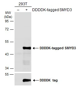
Non-transfected (–) and transfected (+) 293T whole cell extracts (30 μg) were separated by 10% SDS-PAGE, and the membrane was blotted with SMYD3 antibody (GTX121945) diluted at 1:1000. The HRP-conjugated anti-rabbit IgG antibody (GTX213110-01) was used to detect the primary antibody.
 were separated by 10% SDS-PAGE, and the membrane was blotted with SMYD3 antibody (GTX121945) diluted at a dilution of 1:1000.")
 was separated by 10% SDS-PAGE, and the membrane was blotted with SMYD3 antibody (GTX121945) diluted at 1:1000. The HRP-conjugated anti-rabbit IgG antibody (GTX213110-01) was used to detect the primary antibody.")


 were separated by 10% SDS-PAGE, and the membrane was blotted with SMYD3 antibody (GTX121945) diluted at 1:1000. The HRP-conjugated anti-rabbit IgG antibody (GTX213110-01) was used to detect the primary antibody.")
![SMYD3 antibody detects SMYD3 protein by immunofluorescent analysis. Sample: MCF-7 cells were fixed in 4% paraformaldehyde at RT for 15 min. Green: SMYD3 stained by SMYD3 antibody (GTX121945) diluted at 1:500. Red: alpha Tubulin, a cytoskeleton marker, stained by alpha Tubulin antibody [GT114] (GTX628802) diluted at 1:1000. Blue: Fluoroshield with DAPI (GTX30920).](https://www.genetex.com/upload/website/prouct_img/normal/GTX121945/GTX121945_45159_20241213_ICC_IF_24123021_927.webp "SMYD3 antibody detects SMYD3 protein by immunofluorescent analysis. Sample: MCF-7 cells were fixed in 4% paraformaldehyde at RT for 15 min. Green: SMYD3 stained by SMYD3 antibody (GTX121945) diluted at 1:500. Red: alpha Tubulin, a cytoskeleton marker, stained by alpha Tubulin antibody [GT114] (GTX628802) diluted at 1:1000. Blue: Fluoroshield with DAPI (GTX30920).")
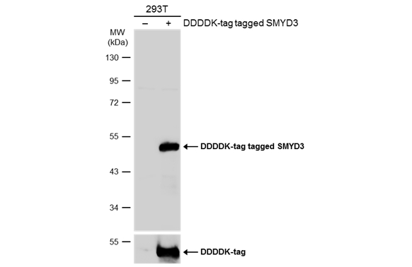
Non-transfected (–) and transfected (+) 293T whole cell extracts (30 μg) were separated by 10% SDS-PAGE, and the membrane was blotted with SMYD3 antibody (GTX121945) diluted at 1:1000. The HRP-conjugated anti-rabbit IgG antibody (GTX213110-01) was used to detect the primary antibody.
SMYD3 antibody
GTX121945
ApplicationsImmunoFluorescence, ImmunoPrecipitation, Western Blot, ImmunoCytoChemistry
Product group Antibodies
ReactivityHuman, Mouse
TargetSMYD3
Overview
- SupplierGeneTex
- Product NameSMYD3 antibody
- Delivery Days Customer9
- Application Supplier NoteWB: 1:500-1:3000. ICC/IF: 1:100-1:1000. IP: 1:100-1:500. *Optimal dilutions/concentrations should be determined by the researcher.Not tested in other applications.
- ApplicationsImmunoFluorescence, ImmunoPrecipitation, Western Blot, ImmunoCytoChemistry
- CertificationResearch Use Only
- ClonalityPolyclonal
- Concentration1.23 mg/ml
- ConjugateUnconjugated
- Gene ID64754
- Target nameSMYD3
- Target descriptionSET and MYND domain containing 3
- Target synonymsKMT3E, ZMYND1, ZNFN3A1, bA74P14.1, histone-lysine N-methyltransferase SMYD3, SET and MYND domain-containing protein 3, zinc finger MYND domain-containing protein 1, zinc finger protein, subfamily 3A (MYND domain containing), 1, zinc finger, MYND domain containing 1
- HostRabbit
- IsotypeIgG
- Protein IDQ9H7B4
- Protein NameHistone-lysine N-methyltransferase SMYD3
- Scientific DescriptionSMYD3 is a histone methyltransferase that plays a role in transcriptional regulation as a member of an RNA polymerase complex.[supplied by OMIM]
- ReactivityHuman, Mouse
- Storage Instruction-20°C or -80°C,2°C to 8°C
- UNSPSC41116161
Datasheet
Related products
Product group Antibodies
SMYD3 AntibodyCSB-PA076745
ApplicationsELISA, ImmunoHistoChemistry
ReactivityHuman, Mouse
TargetSMYD3
- SizePrice
Product group Antibodies

Anti-SMYD3 [RAB-C231]Ab01885-1.1
ApplicationsFlow Cytometry, ImmunoFluorescence, ImmunoPrecipitation
ReactivityHuman
TargetSMYD3
- SizePrice
Product group Antibodies
Anti-SMYD3 AntibodyA31995
ApplicationsImmunoFluorescence, Western Blot, ImmunoHistoChemistry
ReactivityHuman, Mouse
- SizePrice
Product group Antibodies

SMYD3 AntibodyLS-C749520
ApplicationsWestern Blot
ReactivityHuman, Mouse
TargetSMYD3
- SizePrice

Product group Antibodies
Anti-SMYD3 AntibodyHPA045821
ApplicationsImmunoCytoChemistry, ImmunoHistoChemistry
ReactivityHuman
TargetSMYD3
- SizePrice
Product group Antibodies
SMYD3 Recombinant Antibody, AbBy Fluor-594 ConjugatedBSM-61686R-BF594
ApplicationsFlow Cytometry, ImmunoFluorescence, Western Blot
ReactivityHuman, Mouse, Rat
TargetSMYD3
- SizePrice
![Whole cell extract (30 μg) was separated by 10% SDS-PAGE, and the membrane was blotted with SMYD3 antibody [HL2464] (GTX638782) diluted at 1:1000. The HRP-conjugated anti-rabbit IgG antibody (GTX213110-01) was used to detect the primary antibody, and the signal was developed with Trident ECL plus-Enhanced.](https://www.genetex.com/upload/website/prouct_img/normal/GTX638782/GTX638782_T-45089_20230707_WB_M_23071300_455.webp)
Product group Antibodies
SMYD3 antibody [HL2464]GTX638782
ApplicationsImmunoFluorescence, Western Blot, ImmunoCytoChemistry, ImmunoHistoChemistry, ImmunoHistoChemistry Paraffin
ReactivityHuman, Mouse, Rat, Zebra Fish
TargetSMYD3
- SizePrice
Product group Antibodies
SMYD3 antibodyGTX121956
ApplicationsImmunoPrecipitation, Western Blot, ChIP Chromatin ImmunoPrecipitation, ImmunoHistoChemistry, ImmunoHistoChemistry Paraffin
ReactivityHuman
TargetSMYD3
- SizePrice
Product group Antibodies
Anti-SMYD3 Antibody Picoband(r)PB9893-CARRIER-FREE
ApplicationsWestern Blot, ImmunoHistoChemistry
ReactivityBovine, Canine, Equine, Human, Monkey
TargetSMYD3
- SizePrice
Product group Antibodies
Anti-SMYD3 Antibody144-60690
ApplicationsWestern Blot
ReactivityHuman, Mouse
TargetSMYD3
- SizePrice