

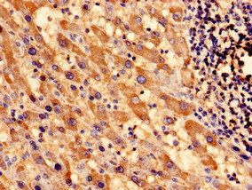
Immunocytochemistry analysis of human liver cancer using CSB-PA022392EA01HU at dilution of 1:100
")
Immunocytochemistry analysis of human liver cancer using CSB-PA022392EA01HU at dilution of 1:100
SOCS3 Antibody
CSB-PA022392EA01HU
ApplicationsImmunoFluorescence, ELISA, ImmunoHistoChemistry
Product group Antibodies
ReactivityHuman
TargetSOCS3
Overview
- SupplierCusabio
- Product NameSOCS3 Antibody
- Delivery Days Customer20
- ApplicationsImmunoFluorescence, ELISA, ImmunoHistoChemistry
- CertificationResearch Use Only
- ClonalityPolyclonal
- ConjugateUnconjugated
- Gene ID9021
- Target nameSOCS3
- Target descriptionsuppressor of cytokine signaling 3
- Target synonymsATOD4, CIS3, Cish3, SOCS-3, SSI-3, SSI3, suppressor of cytokine signaling 3, STAT-induced STAT inhibitor 3, cytokine-inducible SH2 protein 3
- HostRabbit
- IsotypeIgG
- Protein IDO14543
- Protein NameSuppressor of cytokine signaling 3
- Scientific DescriptionSOCS family proteins form part of a classical negative feedback system that regulates cytokine signal transduction. SOCS3 is involved in negative regulation of cytokines that signal through the JAK/STAT pathway. Inhibits cytokine signal transduction by binding to tyrosine kinase receptors including gp130, LIF, erythropoietin, insulin, IL12, GCSF and leptin receptors. Binding to JAK2 inhibits its kinase activity. Suppresses fetal liver erythropoiesis. Regulates onset and maintenance of allergic responses mediated by T-helper type 2 cells. Regulates IL-6 signaling in vivo. Probable substrate recognition component of a SCF-like ECS (Elongin BC-CUL2/5-SOCS-box protein) E3 ubiquitin-protein ligase complex which mediates the ubiquitination and subsequent proteasomal degradation of target proteins. Seems to recognize IL6ST.
- ReactivityHuman
- Storage Instruction-20°C or -80°C
- UNSPSC41116161
Related products

Product group Antibodies
Anti-SOCS-3 AntibodyA96724
ApplicationsWestern Blot, ELISA
ReactivityHuman, Mouse, Rat
- SizePrice
Product group Antibodies
Anti-Mouse SOCS3 Antibody144-00769
ApplicationsWestern Blot
ReactivityHuman, Mouse, Rat
TargetSOCS3
- SizePrice

Product group Antibodies
Anti-SOCS3 Antibody Picoband(r)A00274-2-CARRIER-FREE
ApplicationsFlow Cytometry, Western Blot, ELISA
ReactivityHuman, Mouse, Rat
TargetSOCS3
- SizePrice
Product group Antibodies
SOCS3 Polyclonal AntibodyBS-0580R
ApplicationsImmunoFluorescence, ELISA, ImmunoCytoChemistry, ImmunoHistoChemistry, ImmunoHistoChemistry Frozen, ImmunoHistoChemistry Paraffin
ReactivityBovine, Canine, Equine, Human, Mouse, Porcine, Rabbit, Rat, Sheep
TargetSOCS3
- SizePrice
Product group Antibodies
Socs3 Polyclonal AntibodyCAC08863
ApplicationsImmunoFluorescence, ELISA, ImmunoHistoChemistry
TargetSOCS3
- SizePrice
Product group Antibodies

SOCS3 AntibodyLS-C331075
ApplicationsWestern Blot
ReactivityHuman, Mouse, Rat
TargetSOCS3
- SizePrice

Product group Antibodies
Anti-SOCS3 AntibodyHPA068569
ApplicationsImmunoCytoChemistry
ReactivityHuman
TargetSOCS3
- SizePrice
![Untreated (–) and treated (+) Raw264.7 whole cell extracts (30 μg) were separated by 12% SDS-PAGE, and the membrane was blotted with SOCS3 antibody [C2C3], C-term (GTX104720) diluted at 1:500. The HRP-conjugated anti-rabbit IgG antibody (GTX213110-01) was used to detect the primary antibody, and the signal was developed with Trident ECL plus-Enhanced.](https://www.genetex.com/upload/website/prouct_img/normal/GTX104720/GTX104720_43782_20191213_WB_M_treatment_LPS_w_23060120_855.webp)
Product group Antibodies
SOCS3 antibody [C2C3], C-termGTX104720
ApplicationsWestern Blot
ReactivityHuman, Mouse
TargetSOCS3
- SizePrice