

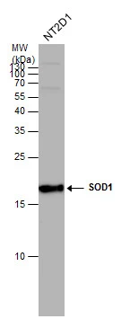
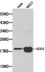
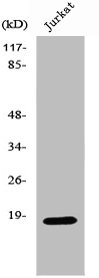
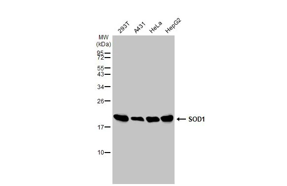
SOD1 antibody detects SOD1 protein by western blot analysis. Whole cell extracts (30 μg) was separated by 15% SDS-PAGE, and the membrane was blotted with SOD1 antibody (GTX100554) at a dilution of 1:1000. The HRP-conjugated anti-rabbit IgG antibody (GTX213110-01) was used to detect the primary antibody.
![Immunofluorescence photomicrographs of paraffin-embedded mouse fetal brain. Green: SOD1 antibody (GTX100554) diluted at 1:200. The signal was developed using goat anti-rabbit IgG antibody (Dylight488) (GTX213110-04). Red: beta Tubulin 3/ TUJ1 antibody [GT11710] diluted at 1:100. The signal was developed using goat anti-mouse IgG antibody (Dylight594) (GTX213111-05). Blue: Nuclear staining with Hoechst 33342.
Antigen Retrieval: Citrate buffer, pH 6.0, 15 min](https://www.genetex.com/upload/website/prouct_img/normal/GTX100554/GTX100554_40163_20150414_IHC-P_M_w_23060100_208.webp "Immunofluorescence photomicrographs of paraffin-embedded mouse fetal brain. Green: SOD1 antibody (GTX100554) diluted at 1:200. The signal was developed using goat anti-rabbit IgG antibody (Dylight488) (GTX213110-04). Red: beta Tubulin 3/ TUJ1 antibody [GT11710] diluted at 1:100. The signal was developed using goat anti-mouse IgG antibody (Dylight594) (GTX213111-05). Blue: Nuclear staining with Hoechst 33342.
Antigen Retrieval: Citrate buffer, pH 6.0, 15 min")
 A: Rat brain 15% SDS PAGE GTX100554 diluted at 1:1000 The HRP-conjugated anti-rabbit IgG antibody (GTX213110-01) was used to detect the primary antibody.")
 were separated by 15% SDS-PAGE, and the membrane was blotted with SOD1 antibody (GTX100554) diluted at 1:1000. The HRP-conjugated anti-rabbit IgG antibody (GTX213110-01) was used to detect the primary antibody.")
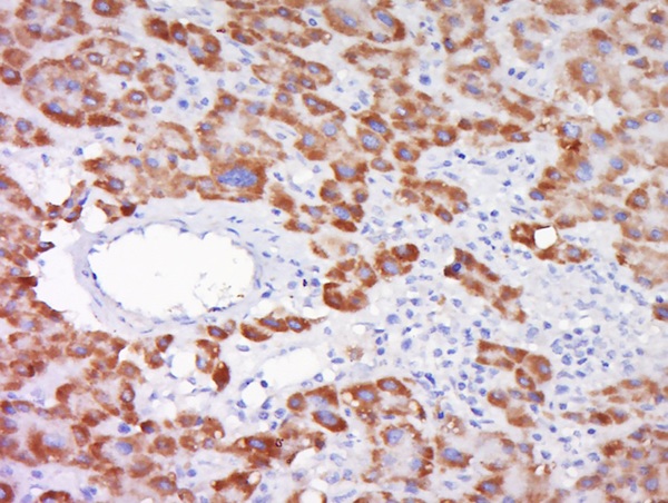
 dilution: 1:500.
Antigen Retrieval: Trilogy? (EDTA based, pH 8.0) buffer, 15min")
 A: mouse brain 15% SDS PAGE GTX100554 diluted at 1:1000 The HRP-conjugated anti-rabbit IgG antibody (GTX213110-01) was used to detect the primary antibody.")

 and transfected (+) 293T whole cell extracts (30 μg) were separated by 15% SDS-PAGE, and the membrane was blotted with SOD1 antibody (GTX100554) diluted at 1:5000. The HRP-conjugated anti-rabbit IgG antibody (GTX213110-01) was used to detect the primary antibody.")


SOD1 antibody detects SOD1 protein by western blot analysis. Whole cell extracts (30 μg) was separated by 15% SDS-PAGE, and the membrane was blotted with SOD1 antibody (GTX100554) at a dilution of 1:1000. The HRP-conjugated anti-rabbit IgG antibody (GTX213110-01) was used to detect the primary antibody.
SOD1 antibody
GTX100554
ApplicationsImmunoFluorescence, Western Blot, ELISA, ImmunoCytoChemistry, ImmunoHistoChemistry, ImmunoHistoChemistry Paraffin
Product group Antibodies
ReactivityAvian, Human, Mouse, Reptile, Rat
TargetSOD1
Overview
- SupplierGeneTex
- Product NameSOD1 antibody
- Delivery Days Customer9
- Application Supplier NoteWB: 1:500-1:10000. ICC/IF: 1:100-1:1000. IHC-P: 1:100-1:1000. ELISA: 1:1000-1:10000. *Optimal dilutions/concentrations should be determined by the researcher.Not tested in other applications.
- ApplicationsImmunoFluorescence, Western Blot, ELISA, ImmunoCytoChemistry, ImmunoHistoChemistry, ImmunoHistoChemistry Paraffin
- CertificationResearch Use Only
- ClonalityPolyclonal
- Concentration0.15 mg/ml
- ConjugateUnconjugated
- Gene ID6647
- Target nameSOD1
- Target descriptionsuperoxide dismutase 1
- Target synonymsALS, ALS1, HEL-S-44, IPOA, SOD, STAHP, hSod1, homodimer, superoxide dismutase [Cu-Zn], Cu/Zn superoxide dismutase, SOD, soluble, epididymis secretory protein Li 44, indophenoloxidase A, superoxide dismutase 1, soluble, superoxide dismutase, cystolic
- HostRabbit
- IsotypeIgG
- Protein IDP00441
- Protein NameSuperoxide dismutase [Cu-Zn]
- Scientific DescriptionThe protein encoded by this gene binds copper and zinc ions and is one of two isozymes responsible for destroying free superoxide radicals in the body. The encoded isozyme is a soluble cytoplasmic protein, acting as a homodimer to convert naturally-occuring but harmful superoxide radicals to molecular oxygen and hydrogen peroxide. The other isozyme is a mitochondrial protein. Mutations in this gene have been implicated as causes of familial amyotrophic lateral sclerosis. Rare transcript variants have been reported for this gene. [provided by RefSeq]
- ReactivityAvian, Human, Mouse, Reptile, Rat
- Storage Instruction-20°C or -80°C,2°C to 8°C
- UNSPSC41116161
Datasheet
Related products
Product group Antibodies
Anti-SOD1 AntibodyA28750
ApplicationsWestern Blot, ImmunoHistoChemistry
ReactivityHuman, Mouse, Rat
- SizePrice
Product group Antibodies
Anti-SOD1 Antibody130-10366
ApplicationsWestern Blot, ELISA
TargetSOD1
- SizePrice
Product group Antibodies

ApplicationsWestern Blot, ELISA
ReactivityHuman
TargetSOD1
- SizePrice
Product group Antibodies
References
SOD1 Polyclonal antibodyBS-10216R
ApplicationsFlow Cytometry, ImmunoFluorescence, Western Blot, ELISA, ImmunoCytoChemistry, ImmunoHistoChemistry, ImmunoHistoChemistry Frozen, ImmunoHistoChemistry Paraffin
ReactivityBovine, Equine, Human, Mouse, Porcine, Rat
TargetSOD1
- SizePrice
Product group Antibodies
SOD1 AntibodyCSB-PA004131
ApplicationsWestern Blot, ELISA
ReactivityHuman, Mouse, Rat
TargetSOD1
- SizePrice
Product group Antibodies

Goat anti-SOD1, BiotinylatedEB07208-B
ApplicationsWestern Blot, ELISA
ReactivityHuman, Mouse, Rat
TargetSOD1
- SizePrice
Product group Antibodies
Sod1 Polyclonal AntibodyCAC07037
ApplicationsImmunoFluorescence, Western Blot, ELISA, ImmunoHistoChemistry
ReactivityMouse
TargetSOD1
- SizePrice
Product group Antibodies
SOD1 antibody [SOD1/2089]GTX18134
ApplicationsELISA, Other Application
ReactivityHuman
TargetSOD1
- SizePrice
Product group Antibodies
SOD1 antibodyGTX100659
ApplicationsWestern Blot, ImmunoHistoChemistry, ImmunoHistoChemistry Frozen
ReactivityHuman, Mouse, Rat
TargetSOD1
- SizePrice
![ICC/IF analysis of PANC-1 (left) and SKBR-3 (right) cells using GTX83012 SOD1 antibody [6F5]. Green : SOD1 Blue: DRAQ5 fluorescent DNA dye Red: Actin filaments](https://www.genetex.com/upload/website/prouct_img/normal/GTX83012/GTX83012_20170912_ICCIF_1_w_23061322_886.webp)
Product group Antibodies
References
SOD1 antibody [6F5]GTX83012
ApplicationsFlow Cytometry, ImmunoFluorescence, Western Blot, ELISA, ImmunoCytoChemistry
ReactivityHuman, Mouse
TargetSOD1
- SizePrice