

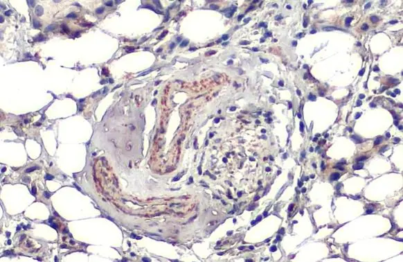
SOD2 antibody [HL1483] detects SOD2 protein at cytoplasm by immunohistochemical analysis. Sample: Paraffin-embedded human breast carcinoma. SOD2 stained by SOD2 antibody [HL1483] (GTX636957) diluted at 1:100. Antigen Retrieval: Citrate buffer, pH 6.0, 15 min
![Non-transfected (–) and transfected (+) 293T whole cell extracts (30 μg) were separated by 12% SDS-PAGE, and the membrane was blotted with SOD2 antibody [HL1483] (GTX636957) diluted at 1:1000. The HRP-conjugated anti-rabbit IgG antibody (GTX213110-01) was used to detect the primary antibody.](https://www.genetex.com/upload/website/prouct_img/normal/GTX636957/GTX636957_T-44669_20220819_WB_B_22082402_219.webp "Non-transfected (–) and transfected (+) 293T whole cell extracts (30 μg) were separated by 12% SDS-PAGE, and the membrane was blotted with SOD2 antibody [HL1483] (GTX636957) diluted at 1:1000. The HRP-conjugated anti-rabbit IgG antibody (GTX213110-01) was used to detect the primary antibody.")
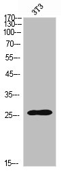
![Various whole cell extracts (30 μg) were separated by 12% SDS-PAGE, and the membrane was blotted with SOD2 antibody [HL1483] (GTX636957) diluted at 1:1000. The HRP-conjugated anti-rabbit IgG antibody (GTX213110-01) was used to detect the primary antibody. Corresponding RNA expression data for the same cell lines are based on Human Protein Atlas program.](https://www.genetex.com/upload/website/prouct_img/normal/GTX636957/GTX636957_44760_20220826_WB_TPM_watermark_22083119_551.webp "Various whole cell extracts (30 μg) were separated by 12% SDS-PAGE, and the membrane was blotted with SOD2 antibody [HL1483] (GTX636957) diluted at 1:1000. The HRP-conjugated anti-rabbit IgG antibody (GTX213110-01) was used to detect the primary antibody. Corresponding RNA expression data for the same cell lines are based on Human Protein Atlas program.")
![Whole zebrafish extract (30 μg) was separated by 12% SDS-PAGE, and the membrane was blotted with SOD2 antibody [HL1483] (GTX636957) diluted at 1:1000. The HRP-conjugated anti-rabbit IgG antibody (GTX213110-01) was used to detect the primary antibody.](https://www.genetex.com/upload/website/prouct_img/normal/GTX636957/GTX636957_44760_20221216_WB_Z_22122018_565.webp "Whole zebrafish extract (30 μg) was separated by 12% SDS-PAGE, and the membrane was blotted with SOD2 antibody [HL1483] (GTX636957) diluted at 1:1000. The HRP-conjugated anti-rabbit IgG antibody (GTX213110-01) was used to detect the primary antibody.")
![Non-transfected (–) and transfected (+) 293T whole cell extracts (30 μg) were separated by 12% SDS-PAGE, and the membrane was blotted with SOD2 antibody [HL1483] (GTX636957) diluted at 1:2000. The HRP-conjugated anti-rabbit IgG antibody (GTX213110-01) was used to detect the primary antibody.](https://www.genetex.com/upload/website/prouct_img/normal/GTX636957/GTX636957_44760_20230728_WB_shRNA_watermark_23073119_606.webp "Non-transfected (–) and transfected (+) 293T whole cell extracts (30 μg) were separated by 12% SDS-PAGE, and the membrane was blotted with SOD2 antibody [HL1483] (GTX636957) diluted at 1:2000. The HRP-conjugated anti-rabbit IgG antibody (GTX213110-01) was used to detect the primary antibody.")
![Whole Japanese medaka extract (30 μg) was separated by 12% SDS-PAGE, and the membrane was blotted with SOD2 antibody [HL1483] (GTX636957) diluted at 1:1000. The HRP-conjugated anti-rabbit IgG antibody (GTX213110-01) was used to detect the primary antibody.](https://www.genetex.com/upload/website/prouct_img/normal/GTX636957/GTX636957_44760_20250815_WB_medaka_25082121_730.webp "Whole Japanese medaka extract (30 μg) was separated by 12% SDS-PAGE, and the membrane was blotted with SOD2 antibody [HL1483] (GTX636957) diluted at 1:1000. The HRP-conjugated anti-rabbit IgG antibody (GTX213110-01) was used to detect the primary antibody.")
SOD2 antibody [HL1483] detects SOD2 protein at cytoplasm by immunohistochemical analysis. Sample: Paraffin-embedded human breast carcinoma. SOD2 stained by SOD2 antibody [HL1483] (GTX636957) diluted at 1:100. Antigen Retrieval: Citrate buffer, pH 6.0, 15 min
SOD2 antibody [HL1483]
GTX636957
ApplicationsWestern Blot, ImmunoHistoChemistry, ImmunoHistoChemistry Paraffin
Product group Antibodies
ReactivityHuman, Zebra Fish
TargetSOD2
Overview
- SupplierGeneTex
- Product NameSOD2 antibody [HL1483]
- Delivery Days Customer9
- Application Supplier NoteWB: 1:500-1:3000. IHC-P: 1:100-1:1000. *Optimal dilutions/concentrations should be determined by the researcher.Not tested in other applications.
- ApplicationsWestern Blot, ImmunoHistoChemistry, ImmunoHistoChemistry Paraffin
- CertificationResearch Use Only
- ClonalityMonoclonal
- Clone IDHL1483
- Concentration1 mg/ml
- ConjugateUnconjugated
- Gene ID6648
- Target nameSOD2
- Target descriptionsuperoxide dismutase 2
- Target synonymsGC1, GClnc1, IPO-B, IPOB, MNSOD, MVCD6, Mn-SOD, lncRNA-GC1, superoxide dismutase [Mn], mitochondrial, Mn superoxide dismutase, epididymis secretory sperm binding protein, gastric cancer-associated lncRNA 1, indophenoloxidase B, manganese-containing superoxide dismutase, mangano-superoxide dismutase, superoxide dismutase 2, mitochondrial
- HostRabbit
- IsotypeIgG
- Protein IDP04179
- Protein NameSuperoxide dismutase [Mn], mitochondrial
- Scientific DescriptionThis gene is a member of the iron/manganese superoxide dismutase family. It encodes a mitochondrial protein that forms a homotetramer and binds one manganese ion per subunit. This protein binds to the superoxide byproducts of oxidative phosphorylation and converts them to hydrogen peroxide and diatomic oxygen. Mutations in this gene have been associated with idiopathic cardiomyopathy (IDC), premature aging, sporadic motor neuron disease, and cancer. Alternative splicing of this gene results in multiple transcript variants. A related pseudogene has been identified on chromosome 1. [provided by RefSeq, Apr 2016]
- ReactivityHuman, Zebra Fish
- Storage Instruction-20°C or -80°C,2°C to 8°C
- UNSPSC41116161
Datasheet
Related products
Product group Antibodies
SOD2 AntibodyCSB-PA006154
ApplicationsWestern Blot, ELISA
ReactivityHuman, Mouse, Rat
TargetSOD2
- SizePrice

Product group Antibodies
Anti-SOD2 AntibodyA97904
ApplicationsWestern Blot, ELISA
ReactivityHuman, Mouse, Rat
- SizePrice
Product group Antibodies

Goat anti-MNSOD (isoform A)EB10971
ApplicationsWestern Blot, ELISA, ImmunoHistoChemistry
ReactivityBovine, Human, Porcine
TargetSOD2
- SizePrice

Product group Antibodies
Anti-SOD2 AntibodyHPA001814
ApplicationsWestern Blot, ImmunoHistoChemistry
ReactivityHuman, Mouse, Rat
TargetSOD2
- SizePrice
Product group Antibodies

SOD2 / Mn SOD AntibodyLS-C401040
ApplicationsWestern Blot, ELISA
ReactivityHuman, Mouse, Rat
TargetSOD2
- SizePrice

Product group Antibodies
Anti-SOD2 Antibody Picoband(r)PB9442-CARRIER-FREE
ApplicationsWestern Blot, ImmunoCytoChemistry, ImmunoHistoChemistry
ReactivityHamster, Human, Mouse, Rat
TargetSOD2
- SizePrice
![Non-transfected (–) and transfected (+) 293T whole cell extracts (30 μg) were separated by 12% SDS-PAGE, and the membrane was blotted with SOD2 (Acetyl Lys68) antibody [HL1460] (GTX636934) diluted at 1:5000. The HRP-conjugated anti-rabbit IgG antibody (GTX213110-01) was used to detect the primary antibody.](https://www.genetex.com/upload/website/prouct_img/normal/GTX636934/GTX636934_44788_20220902_WB_B_22092119_512.webp)
Product group Antibodies
ApplicationsDot Blot, Western Blot
ReactivityHuman, Mouse
TargetSOD2
- SizePrice
![WB analysis of various samples using GTX09013 SOD2 antibody [GT1165]. Dilution : 1:1000 Loading : 25 μg](https://www.genetex.com/upload/website/prouct_img/normal/GTX09013/GTX09013_20200508_WB_w_23053123_761.webp)
Product group Antibodies
SOD2 antibody [GT1165]GTX09013
ApplicationsImmunoFluorescence, Western Blot, ImmunoCytoChemistry, ImmunoHistoChemistry, ImmunoHistoChemistry Paraffin
ReactivityHuman, Mouse, Rat
TargetSOD2
- SizePrice