

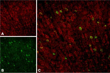
IHC-Fr analysis of rat neocortex tissue using GTX54854 Sortilin 1 antibody. Panel A : Sortilin appears in cortical neurons (red). Panel B : Staining of interneurons with mouse anti-parvalbumin (PV, green). Panel C : Merge of Sortilin and PV demonstrates separate localization in neocortex.

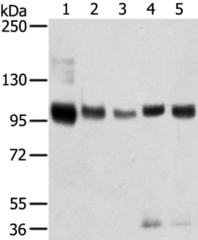
 and rat (lanes 2 and 4) brain lysates using GTX54854 Sortilin 1 antibody preincubated with or without immunogen peptide. Dilution : 1:400")
IHC-Fr analysis of rat neocortex tissue using GTX54854 Sortilin 1 antibody. Panel A : Sortilin appears in cortical neurons (red). Panel B : Staining of interneurons with mouse anti-parvalbumin (PV, green). Panel C : Merge of Sortilin and PV demonstrates separate localization in neocortex.
Sortilin 1 antibody
GTX54854
ApplicationsFlow Cytometry, Western Blot, ImmunoHistoChemistry, ImmunoHistoChemistry Frozen
Product group Antibodies
ReactivityHuman, Mouse, Rat
TargetSORT1
Overview
- SupplierGeneTex
- Product NameSortilin 1 antibody
- Delivery Days Customer7
- ApplicationsFlow Cytometry, Western Blot, ImmunoHistoChemistry, ImmunoHistoChemistry Frozen
- CertificationResearch Use Only
- ClonalityPolyclonal
- Concentration0.4 mg/ml
- ConjugateUnconjugated
- Gene ID6272
- Target nameSORT1
- Target descriptionsortilin 1
- Target synonymsGp95, LDLCQ6, NT3, NTR3, sortilin, 100 kDa NT receptor, glycoprotein 95, neurotensin receptor 3
- HostRabbit
- IsotypeIgG
- Protein IDQ99523
- Protein NameSortilin
- Scientific DescriptionThis gene encodes a member of the VPS10-related sortilin family of proteins. The encoded preproprotein is proteolytically processed by furin to generate the mature receptor. This receptor plays a role in the trafficking of different proteins to either the cell surface, or subcellular compartments such as lysosomes and endosomes. Expression levels of this gene may influence the risk of myocardial infarction in human patients. Alternative splicing results in multiple transcript variants. [provided by RefSeq, Oct 2015]
- ReactivityHuman, Mouse, Rat
- Storage Instruction-20°C or -80°C,2°C to 8°C
- UNSPSC41116161
References
- Phosphatidylinositol (3,4,5)-trisphosphate binds to sortilin and competes with neurotensin: Implications for very low density lipoprotein binding. Sparks RP et al., 2016 Oct 21, Biochem Biophys Res CommunRead this paper
- Sortilin facilitates VLDL-B100 secretion by insulin sensitive McArdle RH7777 cells. Sparks RP et al., 2016 Sep 16, Biochem Biophys Res CommunRead this paper
Datasheet
Related products
Product group Antibodies
Anti-SORT1 AntibodyA38191
ApplicationsWestern Blot, ImmunoHistoChemistry
ReactivityHuman, Mouse, Rat
- SizePrice
![Binding of Progranulin (human) (rec.) (untagged) (Prod. No. AG-40A-0188Y) to Sortilin (human) (rec.) (His) (Prod. No. AG-40B-0229) is inhibited by the antibody Sortilin (human), mAb (rec.) (blocking) [Latozinemab Bioimilar] (Prod. No. AG-27B-7000PF). Methods: Progranulin (human) (rec.) (untagged) is coated on an ELISA plate at 1 microg/ml. Sortilin (human), mAb (rec.) (blocking) [Latozinemab Biosimilar] or an unrelated mAb (Control) is added (starting at 20 microg/ml with a twofold serial dilution) together with 250 ng/ml of Sortilin (rec.) (His) for 1 hour. The binding is detected using an anti-His (HRP) incubated for 30 minutes followed by addition of the substrate TMB. The decreasing ODs observed in the Y axis represent the binding between Progranulin and Sortilin that is inhibited in a dose-dependent manner by Sortilin (human), mAb (rec.) (blocking) [Latozinemab Biosimilar]. This inhibition is not observed with the control antibody.](https://adipogen.com/pub/media/catalog/product/a/g/ag-27b-7000pf-binding_blocking_assay-500px.jpg)
Product group Antibodies
ApplicationsNeutralisation/Blocking
ReactivityHuman
TargetSORT1
- SizePrice
Product group Antibodies
Anti-SORT1 Antibody144-07926
ApplicationsWestern Blot, ImmunoHistoChemistry
ReactivityHuman, Mouse, Rat
TargetSORT1
- SizePrice

Product group Antibodies
Anti-SORT1 AntibodyAMAB91427
ApplicationsWestern Blot, ImmunoCytoChemistry, ImmunoHistoChemistry
ReactivityHuman, Mouse
TargetSORT1
- SizePrice
Product group Antibodies
NTR3 Polyclonal AntibodyBS-6329R
ApplicationsFlow Cytometry, ImmunoFluorescence, Western Blot, ELISA, ImmunoCytoChemistry, ImmunoHistoChemistry, ImmunoHistoChemistry Frozen, ImmunoHistoChemistry Paraffin
ReactivityBovine, Canine, Equine, Guinea Pig, Human, Mouse, Porcine, Rat
TargetSORT1
- SizePrice
Product group Antibodies
ApplicationsImmunoPrecipitation, Western Blot, ImmunoCytoChemistry, ImmunoHistoChemistry
ReactivityMouse, Porcine, Rat
TargetSORT1
- SizePrice
Product group Antibodies
SORT1 AntibodyCSB-PA194090
ApplicationsELISA, ImmunoHistoChemistry
ReactivityHuman, Mouse, Rat
TargetSORT1
- SizePrice

Product group Antibodies
ApplicationsWestern Blot, ImmunoHistoChemistry
ReactivityHuman
TargetSORT1
- SizePrice
Product group Antibodies

SORT1 / Sortilin AntibodyLS-C403604
ApplicationsWestern Blot, ELISA, ImmunoHistoChemistry
ReactivityHuman, Mouse, Rat
TargetSORT1
- SizePrice
![Sortilin 1 antibody [C1C3] detects Sortilin 1 protein by immunofluorescent analysis. Sample: DIV10 rat E18 primary cortical neurons were fixed in 4% paraformaldehyde at RT for 15 min. Green: Sortilin 1 protein stained by Sortilin 1 antibody [C1C3] (GTX110857) diluted at 1:500. Red: beta Tubulin 3/ Tuj1, stained by beta Tubulin 3/ Tuj1 antibody [GT1338] (GTX631831) diluted at 1:500. Blue: Fluoroshield with DAPI (GTX30920).](https://www.genetex.com/upload/website/prouct_img/normal/GTX110857/GTX110857_40051_20170824_IFA_w_23060500_433.webp)
Product group Antibodies
Sortilin 1 antibody [C1C3]GTX110857
ApplicationsImmunoFluorescence, Western Blot, ImmunoCytoChemistry, ImmunoHistoChemistry, ImmunoHistoChemistry Paraffin
ReactivityHuman, Mouse, Rat
TargetSORT1
- SizePrice