

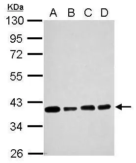
Sample (30 ug of whole cell lysate) A: NT2D1 B: PC-3 C: U87-MG D: SK-N-SH 10% SDS PAGE GTX627404 diluted at 1:5000
![Non-transfected (–) and transfected (+) NT2D1 whole cell extracts (30 μg) were separated by 10% SDS-PAGE, and the membrane was blotted with SOX2 antibody [GT1876] (GTX627404) diluted at 1:500.](https://www.genetex.com/upload/website/prouct_img/normal/GTX627404/GTX627404_40846_20170126_WB_shRNA_watermark_w_23061202_647.webp "Non-transfected (–) and transfected (+) NT2D1 whole cell extracts (30 μg) were separated by 10% SDS-PAGE, and the membrane was blotted with SOX2 antibody [GT1876] (GTX627404) diluted at 1:500.")
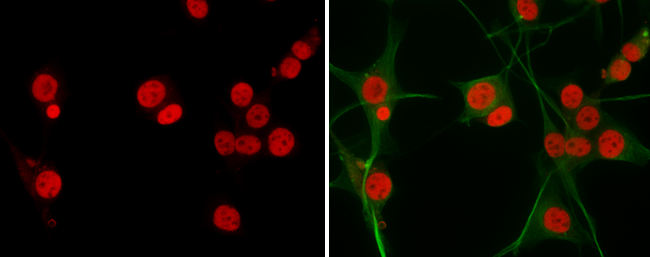
![SOX2 antibody [GT1876] detects SOX2 protein expression at nucleus by immunohistochemical analysis. Sample: Frozen sectioned E13.5 Rat brain. Red: SOX2 protein stained by SOX2 antibody [GT1876] (GTX627404) diluted at 1:250. Blue: Fluoroshield with DAPI (GTX30920).](https://www.genetex.com/upload/website/prouct_img/normal/GTX627404/GTX627404_40846_20160921_IHC-Fr_w_23061202_430.webp "SOX2 antibody [GT1876] detects SOX2 protein expression at nucleus by immunohistochemical analysis. Sample: Frozen sectioned E13.5 Rat brain. Red: SOX2 protein stained by SOX2 antibody [GT1876] (GTX627404) diluted at 1:250. Blue: Fluoroshield with DAPI (GTX30920).")
![SOX2 antibody [GT1876] detects SOX2 protein by immunohistochemical analysis. Sample: Frozen sectioned adult mouse retina. Green: SOX2 protein stained by SOX2 antibody [GT1876] (GTX627404) diluted at 1:250. Red: PKC alpha protein stained by PKC alpha antibody [GT1876] (GTX130453) diluted at 1:250. Blue: Fluoroshield with DAPI (GTX30920).](https://www.genetex.com/upload/website/prouct_img/normal/GTX627404/GTX627404_40846_20160824_IHC-Fr_w_23061202_235.webp "SOX2 antibody [GT1876] detects SOX2 protein by immunohistochemical analysis. Sample: Frozen sectioned adult mouse retina. Green: SOX2 protein stained by SOX2 antibody [GT1876] (GTX627404) diluted at 1:250. Red: PKC alpha protein stained by PKC alpha antibody [GT1876] (GTX130453) diluted at 1:250. Blue: Fluoroshield with DAPI (GTX30920).")
![Whole cell extract (30 μg) was separated by 12% SDS-PAGE, and the membranes were blotted with SOX2 antibody [GT1876] (GTX627404) diluted at 1:1000 and competitor's antibody (sc-17320) diluted at 1:500. The HRP-conjugated anti-mouse IgG antibody (GTX213111-01) was used to detect the primary antibody. *The competitor is not affiliated with GeneTex and does not endorse this product.](https://www.genetex.com/upload/website/prouct_img/normal/GTX627404/GTX627404_40846_20180119_WB_competitor_watermark_w_23061202_638.webp "Whole cell extract (30 μg) was separated by 12% SDS-PAGE, and the membranes were blotted with SOX2 antibody [GT1876] (GTX627404) diluted at 1:1000 and competitor's antibody (sc-17320) diluted at 1:500. The HRP-conjugated anti-mouse IgG antibody (GTX213111-01) was used to detect the primary antibody. *The competitor is not affiliated with GeneTex and does not endorse this product.")
![SOX2 antibody [GT1876] detects SOX2 protein by Western blot analysis. A. 30 μg zebrafish eye lysate/extract 10 % SDS-PAGE SOX2 antibody [GT1876] (GTX627404) dilution: 1:500](https://www.genetex.com/upload/website/prouct_img/normal/GTX627404/GTX627404_40846_WB_Z_eye_w_23061202_549.webp "SOX2 antibody [GT1876] detects SOX2 protein by Western blot analysis. A. 30 μg zebrafish eye lysate/extract 10 % SDS-PAGE SOX2 antibody [GT1876] (GTX627404) dilution: 1:500")
![SOX2 antibody [GT1876] detects SOX2 protein by Western blot analysis. A. 30 μg human ESC whole cell lysate/extract 10 % SDS-PAGE SOX2 antibody [GT1876] (GTX627404) dilution: 1:5000](https://www.genetex.com/upload/website/prouct_img/normal/GTX627404/GTX627404_40846_WB_hESC_w_23061202_171.webp "SOX2 antibody [GT1876] detects SOX2 protein by Western blot analysis. A. 30 μg human ESC whole cell lysate/extract 10 % SDS-PAGE SOX2 antibody [GT1876] (GTX627404) dilution: 1:5000")
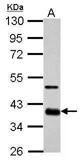
![Immunoprecipitation of SOX2 protein from NT2D1 whole cell extracts using 5 μg of SOX2 antibody [GT1876] (GTX627404) or SOX2 antibody [N1C3] (GTX101057). Western blot analysis was performed using SOX2 antibody [GT1876] (GTX627404) diluted at 1:500. EasyBlot anti-Mouse IgG (GTX221667-01) was used as a secondary reagent.](https://www.genetex.com/upload/website/prouct_img/normal/GTX627404/GTX627404_40846_IP_2_w_23061202_217.webp "Immunoprecipitation of SOX2 protein from NT2D1 whole cell extracts using 5 μg of SOX2 antibody [GT1876] (GTX627404) or SOX2 antibody [N1C3] (GTX101057). Western blot analysis was performed using SOX2 antibody [GT1876] (GTX627404) diluted at 1:500. EasyBlot anti-Mouse IgG (GTX221667-01) was used as a secondary reagent.")
![Sox2 antibodies detects Sox2 proteins on embryonic mouse brain by immunohistochemical analysis. Sample: Frozen section of embryonic mouse brain (mE18.5). Green: GFAP antibody (GTX100850) diluted at 1:500. Red: Sox2 antibody [GT1876] (GTX627404) diluted at 1:500.](https://www.genetex.com/upload/website/prouct_img/normal/GTX627404/GTX627404_40846_20150625_IHC_M_w_23061202_614.webp "Sox2 antibodies detects Sox2 proteins on embryonic mouse brain by immunohistochemical analysis. Sample: Frozen section of embryonic mouse brain (mE18.5). Green: GFAP antibody (GTX100850) diluted at 1:500. Red: Sox2 antibody [GT1876] (GTX627404) diluted at 1:500.")
![SOX2 antibody [GT1876] detects SOX2 protein by flow cytomertry analysis. Sample: Human embryonic stem cells Black: Isotype control dilution: 1:50 Green: SOX2 antibody [GT1876] dilution: 1:50](https://www.genetex.com/upload/website/prouct_img/normal/GTX627404/GTX627404_40846_CT_FACS_w_23061202_358.webp "SOX2 antibody [GT1876] detects SOX2 protein by flow cytomertry analysis. Sample: Human embryonic stem cells Black: Isotype control dilution: 1:50 Green: SOX2 antibody [GT1876] dilution: 1:50")
Sample (30 ug of whole cell lysate) A: NT2D1 B: PC-3 C: U87-MG D: SK-N-SH 10% SDS PAGE GTX627404 diluted at 1:5000
SOX2 antibody [GT1876]
GTX627404
ApplicationsFlow Cytometry, ImmunoFluorescence, ImmunoPrecipitation, Western Blot, ImmunoCytoChemistry, ImmunoHistoChemistry, ImmunoHistoChemistry Frozen, ImmunoHistoChemistry Paraffin
Product group Antibodies
ReactivityHuman, Mouse, Rat, Zebra Fish
TargetSOX2
Overview
- SupplierGeneTex
- Product NameSOX2 antibody [GT1876]
- Delivery Days Customer9
- Application Supplier NoteWB: 1:500-1:10000. IHC-Fr: 1:100-1:1000. FACS: 1:50-1:200. IP: 1:100-1:500. *Optimal dilutions/concentrations should be determined by the researcher.Not tested in other applications.
- ApplicationsFlow Cytometry, ImmunoFluorescence, ImmunoPrecipitation, Western Blot, ImmunoCytoChemistry, ImmunoHistoChemistry, ImmunoHistoChemistry Frozen, ImmunoHistoChemistry Paraffin
- CertificationResearch Use Only
- ClonalityMonoclonal
- Clone IDGT1876
- Concentration1 mg/ml
- ConjugateUnconjugated
- Gene ID6657
- Target nameSOX2
- Target descriptionSRY-box transcription factor 2
- Target synonymsANOP3, MCOPS3, transcription factor SOX-2, SRY (sex determining region Y)-box 2, SRY-box 2, SRY-related HMG-box gene 2, sex determining region Y-box 2, transcription factor SOX2
- HostMouse
- IsotypeIgG1
- Protein IDP48431
- Protein NameTranscription factor SOX-2
- Scientific DescriptionThis intronless gene encodes a member of the SRY-related HMG-box (SOX) family of transcription factors involved in the regulation of embryonic development and in the determination of cell fate. The product of this gene is required for stem-cell maintenance in the central nervous system, and also regulates gene expression in the stomach. Mutations in this gene have been associated with optic nerve hypoplasia and with syndromic microphthalmia, a severe form of structural eye malformation. This gene lies within an intron of another gene called SOX2 overlapping transcript (SOX2OT). [provided by RefSeq]
- ReactivityHuman, Mouse, Rat, Zebra Fish
- Storage Instruction-20°C or -80°C,2°C to 8°C
- UNSPSC12352203
References
- Mashiko T, Kanayama K, Saito N, et al. Selective Proliferation of Highly Functional Adipose-Derived Stem Cells in Microgravity Culture with Stirred Microspheres. Cells. 2021,10(3). doi: 10.3390/cells10030560Read this paper
- Lee KY, Kuo TC, Chou CM, et al. Upregulation of CD109 Promotes the Epithelial-to-Mesenchymal Transition and Stemness Properties of Lung Adenocarcinomas via Activation of the Hippo-YAP Signaling. Cells. 2020,10(1). doi: 10.3390/cells10010028Read this paper
- Zhang L, Liu F, Weygant N, et al. A novel integrated system using patient-derived glioma cerebral organoids and xenografts for disease modeling and drug screening. Cancer Lett. 2021,500:87-97. doi: 10.1016/j.canlet.2020.12.013Read this paper
- Ma L, Lou S, Miao Z, et al. Identification of novel susceptibility loci for non-syndromic cleft lip with or without cleft palate. J Cell Mol Med. 2020,24(23):13669-13678. doi: 10.1111/jcmm.15878Read this paper
- Andreucci E, Peppicelli S, Ruzzolini J, et al. The acidic tumor microenvironment drives a stem-like phenotype in melanoma cells. J Mol Med (Berl). 2020,98(10):1431-1446. doi: 10.1007/s00109-020-01959-yRead this paper
- Aminuddin A, Ng PY, Leong CO, et al. Mitochondrial DNA alterations may influence the cisplatin responsiveness of oral squamous cell carcinoma. Sci Rep. 2020,10(1):7885. doi: 10.1038/s41598-020-64664-3Read this paper
- Peng L, Zhou Y, Xu W, et al. Generation of Stable Induced Pluripotent Stem-like Cells from Adult Zebra Fish Fibroblasts. Int J Biol Sci. 2019,15(11):2340-2349. doi: 10.7150/ijbs.34010Read this paper
- Lin YH, Wu MH, Huang YH, et al. Thyroid hormone negatively regulates tumorigenesis through suppression of BC200. Endocr Relat Cancer. 2018,25(12):967-979. doi: 10.1530/ERC-18-0176Read this paper
- Chuang HM, Ho LI, Huang MH, et al. Non-Canonical Regulation of Type I Collagen through Promoter Binding of SOX2 and Its Contribution to Ameliorating Pulmonary Fibrosis by Butylidenephthalide. Int J Mol Sci. 2018,19(10). doi: 10.3390/ijms19103024Read this paper
- Chang KY, Huang CT, Hsu TI, et al. Stress stimuli induce cancer-stemness gene expression via Sp1 activation leading to therapeutic resistance in glioblastoma. Biochem Biophys Res Commun. 2017,493(1):14-19. doi: 10.1016/j.bbrc.2017.09.095Read this paper
Datasheet
Related products
Product group Antibodies
Anti-SOX2 Antibody130-10236
ApplicationsWestern Blot, ELISA
TargetSOX2
- SizePrice

Product group Antibodies
Anti-SOX2 AntibodyAMAB91307
ApplicationsWestern Blot, ImmunoCytoChemistry, ImmunoHistoChemistry
ReactivityHuman, Mouse
TargetSOX2
- SizePrice

Product group Antibodies
Anti-SOX2 Antibody Picoband(r)A00105-1-CARRIER-FREE
ApplicationsWestern Blot, ELISA
ReactivityHuman, Rat
TargetSOX2
- SizePrice
Product group Antibodies
References
SOX2 antibodyGTX101506
ApplicationsFlow Cytometry, ImmunoFluorescence, Western Blot, ImmunoCytoChemistry, ImmunoHistoChemistry, ImmunoHistoChemistry Frozen
ReactivityHuman, Mouse
TargetSOX2
- SizePrice
Product group Antibodies
References
SOX2 antibody [N1C3]GTX101507
ApplicationsFlow Cytometry, ImmunoFluorescence, ImmunoPrecipitation, Western Blot, ImmunoCytoChemistry, ImmunoHistoChemistry, ImmunoHistoChemistry Frozen, ImmunoHistoChemistry Paraffin
ReactivityAmphibian, Human, Mammals, Mouse, Rat
TargetSOX2
- SizePrice
![ICC/IF analysis of NTERA-2 cells using GTX60381 SOX2 antibody [10F10]. Green : SOX2 Red: Actin filaments](https://www.genetex.com/upload/website/prouct_img/normal/GTX60381/GTX60381_20170912_ICCIF_w_23061123_336.webp)
Product group Antibodies
SOX2 antibody [10F10]GTX60381
ApplicationsImmunoFluorescence, Western Blot, ELISA, ImmunoCytoChemistry, ImmunoHistoChemistry, ImmunoHistoChemistry Paraffin
ReactivityHuman
TargetSOX2
- SizePrice
Product group Antibodies
References
SOX2 antibody [GT1352]GTX627405
ApplicationsFlow Cytometry, ImmunoFluorescence, Western Blot, ImmunoCytoChemistry, ImmunoHistoChemistry, ImmunoHistoChemistry Frozen, ImmunoHistoChemistry Paraffin
ReactivityHuman, Mouse
TargetSOX2
- SizePrice
![Zebrafish tissue extract (50 μg) was separated by 12% SDS-PAGE, and the membrane was blotted with SOX2 antibody [HL1192] (GTX636504) diluted at 1:1000. The HRP-conjugated anti-rabbit IgG antibody (GTX213110-01) was used to detect the primary antibody.](https://www.genetex.com/upload/website/prouct_img/normal/GTX636504/GTX636504_44508_20211203_WB_Z_brain_24112018_555.webp)
Product group Antibodies
SOX2 antibody [HL1192]GTX636504
ApplicationsWestern Blot, ImmunoHistoChemistry
ReactivityHuman, Zebra Fish
TargetSOX2
- SizePrice
![Mouse ESC extract (50 μg) was separated by 12% SDS-PAGE, and the membrane was blotted with SOX2 antibody [HL1193] (GTX636505) diluted at 1:1000. The HRP-conjugated anti-rabbit IgG antibody (GTX213110-01) was used to detect the primary antibody.](https://www.genetex.com/upload/website/prouct_img/normal/GTX636505/GTX636505_T-44452_20230324_WB_M_ESC_23073119_588.webp)
Product group Antibodies
SOX2 antibody [HL1193]GTX636505
ApplicationsImmunoFluorescence, ImmunoPrecipitation, Western Blot, ImmunoCytoChemistry, ImmunoHistoChemistry, ImmunoHistoChemistry Paraffin
ReactivityFeline, Human, Mouse, Rat
TargetSOX2
- SizePrice