

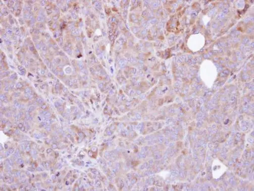
Immunohistochemical analysis of paraffin-embedded SW480 xenograft , using Steroid sulfatase (GTX105498) antibody at 1:100 dilution.
Antigen Retrieval: Trilogy? (EDTA based, pH 8.0) buffer, 15min
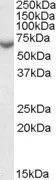
![HeLa whole cell and membrane extracts (30 μg) were separated by 7.5% SDS-PAGE, and the membrane was blotted with Steroid sulfatase antibody [N1C3] (GTX105498) diluted at 1:1000. The HRP-conjugated anti-rabbit IgG antibody (GTX213110-01) was used to detect the primary antibody.](https://www.genetex.com/upload/website/prouct_img/normal/GTX105498/GTX105498_40002_20190426_WB_Fraction_w_23060120_946.webp "HeLa whole cell and membrane extracts (30 μg) were separated by 7.5% SDS-PAGE, and the membrane was blotted with Steroid sulfatase antibody [N1C3] (GTX105498) diluted at 1:1000. The HRP-conjugated anti-rabbit IgG antibody (GTX213110-01) was used to detect the primary antibody.")
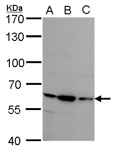
![Human tissue extract (30 μg) was separated by 7.5% SDS-PAGE, and the membrane was blotted with Steroid sulfatase antibody [N1C3] (GTX105498) diluted at 1:500. The HRP-conjugated anti-rabbit IgG antibody (GTX213110-01) was used to detect the primary antibody.](https://www.genetex.com/upload/website/prouct_img/normal/GTX105498/GTX105498_40002_20200424_WB_placenta_w_23060120_641.webp "Human tissue extract (30 μg) was separated by 7.5% SDS-PAGE, and the membrane was blotted with Steroid sulfatase antibody [N1C3] (GTX105498) diluted at 1:500. The HRP-conjugated anti-rabbit IgG antibody (GTX213110-01) was used to detect the primary antibody.")
Immunohistochemical analysis of paraffin-embedded SW480 xenograft , using Steroid sulfatase (GTX105498) antibody at 1:100 dilution.
Antigen Retrieval: Trilogy? (EDTA based, pH 8.0) buffer, 15min
Steroid sulfatase antibody [N1C3]
GTX105498
ApplicationsWestern Blot, ImmunoHistoChemistry, ImmunoHistoChemistry Paraffin
Product group Antibodies
ReactivityHuman
TargetSTS
Overview
- SupplierGeneTex
- Product NameSteroid sulfatase antibody [N1C3]
- Delivery Days Customer9
- Application Supplier NoteWB: 1:500-1:3000. IHC-P: 1:100-1:1000. *Optimal dilutions/concentrations should be determined by the researcher.Not tested in other applications.
- ApplicationsWestern Blot, ImmunoHistoChemistry, ImmunoHistoChemistry Paraffin
- CertificationResearch Use Only
- ClonalityPolyclonal
- Concentration0.43 mg/ml
- ConjugateUnconjugated
- Gene ID412
- Target nameSTS
- Target descriptionsteroid sulfatase
- Target synonymsARSC, ARSC1, ASC, ES, SSDD, XLI, steryl-sulfatase, arylsulfatase C, estrone sulfatase, steroid sulfatase (microsomal), isozyme S, steryl-sulfate sulfohydrolase
- HostRabbit
- IsotypeIgG
- Protein IDP08842
- Protein NameSteryl-sulfatase
- Scientific DescriptionThe protein encoded by this gene catalyzes the conversion of sulfated steroid precursors to estrogens during pregnancy. The encoded protein is found in the endoplasmic reticulum, where it acts as a homodimer. Mutations in this gene are known to cause X-linked ichthyosis (XLI). [provided by RefSeq]
- ReactivityHuman
- Storage Instruction-20°C or -80°C,2°C to 8°C
- UNSPSC41116161
Datasheet
Related products
Product group Antibodies
STS AntibodyCSB-PA022883GA01HU
ApplicationsWestern Blot, ELISA, ImmunoHistoChemistry
ReactivityHuman, Mouse, Rat
TargetSTS
- SizePrice
Product group Antibodies
ApplicationsImmunoPrecipitation, Western Blot, ImmunoCytoChemistry, ImmunoHistoChemistry
ReactivityEquine, Mouse
TargetSTS
- SizePrice

Product group Antibodies
Anti-Steroid sulfatase/STS Antibody Picoband(r)A01198-1-CARRIER-FREE
ApplicationsFlow Cytometry, Western Blot, ELISA
ReactivityHuman, Mouse, Rat
TargetSTS
- SizePrice
Product group Antibodies

ApplicationsWestern Blot, ELISA
ReactivityHuman
TargetSTS
- SizePrice

Product group Antibodies
Anti-STS AntibodyHPA002904
ApplicationsImmunoHistoChemistry
ReactivityHuman
TargetSTS
- SizePrice
Product group Antibodies

ApplicationsWestern Blot
ReactivityHuman, Mouse
TargetSTS
- SizePrice

Product group Antibodies
ApplicationsImmunoFluorescence, Western Blot, ELISA, ImmunoCytoChemistry, ImmunoHistoChemistry, ImmunoHistoChemistry Frozen, ImmunoHistoChemistry Paraffin
ReactivityBovine, Canine, Equine, Human, Mouse, Porcine, Rabbit, Rat
TargetSTS
- SizePrice
Product group Antibodies
Steroid sulfatase antibodyGTX113628
ApplicationsWestern Blot
ReactivityHuman
TargetSTS
- SizePrice
Product group Antibodies
ApplicationsWestern Blot
ReactivityHuman
TargetSTS
- SizePrice
Product group Antibodies
Anti-STS (C-term) Antibody102-21909
ApplicationsWestern Blot, ImmunoHistoChemistry, ImmunoHistoChemistry Paraffin
TargetSTS
- SizePrice