

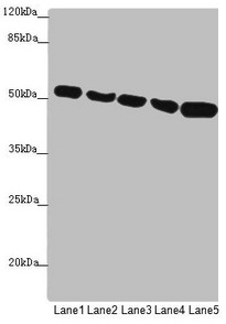
Western blot All lanes: SUCLA2 antibody at 1.75microg/ml Lane 1: Hela whole cell lysate Lane 2: Jurkat whole cell lysate Lane 3: A549 whole cell lysate Lane 4: 293T whole cell lysate Lane 5: HepG2 whole cell lysate Secondary Goat polyclonal to rabbit IgG at 1/10000 dilution Predicted band size: 51, 49 kDa Observed band size: 51 kDa


Western blot All lanes: SUCLA2 antibody at 1.75microg/ml Lane 1: Hela whole cell lysate Lane 2: Jurkat whole cell lysate Lane 3: A549 whole cell lysate Lane 4: 293T whole cell lysate Lane 5: HepG2 whole cell lysate Secondary Goat polyclonal to rabbit IgG at 1/10000 dilution Predicted band size: 51, 49 kDa Observed band size: 51 kDa
SUCLA2 Antibody
CSB-PA868398ESR1HU
ApplicationsWestern Blot, ELISA, ImmunoHistoChemistry
Product group Antibodies
ReactivityHuman
TargetSUCLA2
Overview
- SupplierCusabio
- Product NameSUCLA2 Antibody
- Delivery Days Customer20
- ApplicationsWestern Blot, ELISA, ImmunoHistoChemistry
- CertificationResearch Use Only
- ClonalityPolyclonal
- ConjugateUnconjugated
- Gene ID8803
- Target nameSUCLA2
- Target descriptionsuccinate-CoA ligase ADP-forming subunit beta
- Target synonymsA-BETA, A-SCS, LINC00444, MTDPS5, SCS-betaA, succinate--CoA ligase [ADP-forming] subunit beta, mitochondrial, ATP-specific succinyl-CoA synthetase subunit beta, ATP-specific succinyl-CoA synthetase, beta subunit, long intergenic non-protein coding RNA 444, mitochondrial succinyl-CoA ligase [ADP-forming] subunit beta, renal carcinoma antigen NY-REN-39, succinate-CoA ligase ADP-forming beta subunit, succinate-CoA ligase beta subunit, succinyl-CoA ligase [ADP-forming] subunit beta, mitochondrial, succinyl-CoA synthetase beta-A chain
- HostRabbit
- IsotypeIgG
- Protein IDQ9P2R7
- Protein NameSuccinate--CoA ligase [ADP-forming] subunit beta, mitochondrial
- Scientific DescriptionCatalyzes the ATP-dependent ligation of succinate and CoA to form succinyl-CoA.
- ReactivityHuman
- Storage Instruction-20°C or -80°C
- UNSPSC41116161
Related products

Product group Antibodies
Anti-SUCLA2 Antibody Picoband(r)A04807-1-CARRIER-FREE
ApplicationsFlow Cytometry, ImmunoFluorescence, Western Blot, ELISA, ImmunoCytoChemistry
ReactivityHuman, Mouse, Rat
TargetSUCLA2
- SizePrice

Product group Antibodies
Anti-SUCLA2 AntibodyA12768
ApplicationsImmunoFluorescence, Western Blot, ImmunoCytoChemistry
ReactivityHuman, Mouse, Rat
- SizePrice
Product group Antibodies
Anti-SUCLA2 Antibody144-10040
ApplicationsWestern Blot
ReactivityHuman, Mouse, Rat
TargetSUCLA2
- SizePrice
Product group Antibodies

SUCLA2 AntibodyLS-C496750
ApplicationsWestern Blot
ReactivityHuman, Mouse, Rat
TargetSUCLA2
- SizePrice

Product group Antibodies
Anti-SUCLA2 AntibodyHPA039536
ApplicationsWestern Blot, ImmunoCytoChemistry, ImmunoHistoChemistry
ReactivityHuman
TargetSUCLA2
- SizePrice
![SUCLA2 antibody [N2C3] detects SUCLA2 protein at cytoplasm on mouse stomach by immunohistochemical analysis. Sample: Paraffin-embedded mouse stomach. SUCLA2 antibody [N2C3] (GTX109728) diluted at 1:500.
Antigen Retrieval: Trilogy? (EDTA based, pH 8.0) buffer, 15min](https://www.genetex.com/upload/website/prouct_img/normal/GTX109728/GTX109728_40058_20150226_IHC_M_w_23060500_997.webp)
Product group Antibodies
SUCLA2 antibody [N2C3]GTX109728
ApplicationsImmunoFluorescence, Western Blot, ImmunoCytoChemistry, ImmunoHistoChemistry, ImmunoHistoChemistry Paraffin
ReactivityHuman, Mouse
TargetSUCLA2
- SizePrice