

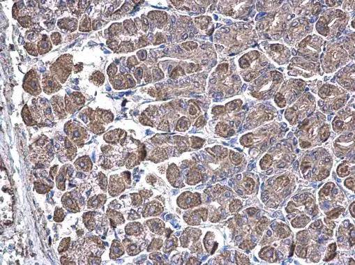
SUCLA2 antibody [N2C3] detects SUCLA2 protein at cytoplasm on mouse stomach by immunohistochemical analysis. Sample: Paraffin-embedded mouse stomach. SUCLA2 antibody [N2C3] (GTX109728) diluted at 1:500.
Antigen Retrieval: Trilogy? (EDTA based, pH 8.0) buffer, 15min
![SUCLA2 antibody [N2C3] detects SUCLA2 protein at mitochondria by immunofluorescent analysis. Sample: HeLa cells were fixed in ice-cold MeOH for 5 min. Green: SUCLA2 protein stained by SUCLA2 antibody [N2C3] (GTX109728) diluted at 1:500. Blue: Hoechst 33342 staining.](https://www.genetex.com/upload/website/prouct_img/normal/GTX109728/GTX109728_40058_20171012_IFA_w_23060500_507.webp "SUCLA2 antibody [N2C3] detects SUCLA2 protein at mitochondria by immunofluorescent analysis. Sample: HeLa cells were fixed in ice-cold MeOH for 5 min. Green: SUCLA2 protein stained by SUCLA2 antibody [N2C3] (GTX109728) diluted at 1:500. Blue: Hoechst 33342 staining.")
 A: mouse brain 10% SDS PAGE GTX109728 diluted at 1:20000")
 antibody at 1:500 dilution.
Antigen Retrieval: Trilogy? (EDTA based, pH 8.0) buffer, 15min")
![Wild-type (WT) and SUCLA2 knockout (KO) HeLa cell extracts (30 μg) were separated by 10% SDS-PAGE, and the membrane was blotted with SUCLA2 antibody [N2C3] (GTX109728) diluted at 1:1000. The HRP-conjugated anti-rabbit IgG antibody (GTX213110-01) was used to detect the primary antibody.](https://www.genetex.com/upload/website/prouct_img/normal/GTX109728/GTX109728_40058_20181123_WB_KO_watermark_w_23060500_140.webp "Wild-type (WT) and SUCLA2 knockout (KO) HeLa cell extracts (30 μg) were separated by 10% SDS-PAGE, and the membrane was blotted with SUCLA2 antibody [N2C3] (GTX109728) diluted at 1:1000. The HRP-conjugated anti-rabbit IgG antibody (GTX213110-01) was used to detect the primary antibody.")
 A: Hep G2 (GTX27900) B: Molt-4 (GTX27912) 10% SDS PAGE GTX109728 diluted at 1:1000")
SUCLA2 antibody [N2C3] detects SUCLA2 protein at cytoplasm on mouse stomach by immunohistochemical analysis. Sample: Paraffin-embedded mouse stomach. SUCLA2 antibody [N2C3] (GTX109728) diluted at 1:500.
Antigen Retrieval: Trilogy? (EDTA based, pH 8.0) buffer, 15min
SUCLA2 antibody [N2C3]
GTX109728
ApplicationsImmunoFluorescence, Western Blot, ImmunoCytoChemistry, ImmunoHistoChemistry, ImmunoHistoChemistry Paraffin
Product group Antibodies
ReactivityHuman, Mouse
TargetSUCLA2
Overview
- SupplierGeneTex
- Product NameSUCLA2 antibody [N2C3]
- Delivery Days Customer9
- Application Supplier NoteWB: 1:500-1:3000. ICC/IF: 1:100-1:1000. IHC-P: 1:100-1:1000. *Optimal dilutions/concentrations should be determined by the researcher.Not tested in other applications.
- ApplicationsImmunoFluorescence, Western Blot, ImmunoCytoChemistry, ImmunoHistoChemistry, ImmunoHistoChemistry Paraffin
- CertificationResearch Use Only
- ClonalityPolyclonal
- Concentration1 mg/ml
- ConjugateUnconjugated
- Gene ID8803
- Target nameSUCLA2
- Target descriptionsuccinate-CoA ligase ADP-forming subunit beta
- Target synonymsA-BETA, A-SCS, LINC00444, MTDPS5, SCS-betaA, succinate--CoA ligase [ADP-forming] subunit beta, mitochondrial, ATP-specific succinyl-CoA synthetase subunit beta, ATP-specific succinyl-CoA synthetase, beta subunit, long intergenic non-protein coding RNA 444, mitochondrial succinyl-CoA ligase [ADP-forming] subunit beta, renal carcinoma antigen NY-REN-39, succinate-CoA ligase ADP-forming beta subunit, succinate-CoA ligase beta subunit, succinyl-CoA ligase [ADP-forming] subunit beta, mitochondrial, succinyl-CoA synthetase beta-A chain
- HostRabbit
- IsotypeIgG
- Protein IDQ9P2R7
- Protein NameSuccinate--CoA ligase [ADP-forming] subunit beta, mitochondrial
- Scientific DescriptionSuccinyl-CoA synthetase (SCS) is a mitochondrial matrix enzyme that acts as a heterodimer, being composed of an invariant alpha subunit and a substrate-specific beta subunit. The protein encoded by this gene is an ATP-specific SCS beta subunit that dimerizes with the SCS alpha subunit to form SCS-A, an essential component of the tricarboxylic acid cycle. SCS-A hydrolyzes ATP to convert succinate to succinyl-CoA. Defects in this gene are a cause of myopathic mitochondrial DNA depletion syndrome. A pseudogene of this gene has been found on chromosome 6. [provided by RefSeq]
- ReactivityHuman, Mouse
- Storage Instruction-20°C or -80°C,2°C to 8°C
- UNSPSC41116161
Datasheet
Related products

Product group Antibodies
Anti-SUCLA2 Antibody Picoband(r)A04807-1-CARRIER-FREE
ApplicationsFlow Cytometry, ImmunoFluorescence, Western Blot, ELISA, ImmunoCytoChemistry
ReactivityHuman, Mouse, Rat
TargetSUCLA2
- SizePrice

Product group Antibodies
Anti-SUCLA2 AntibodyA12768
ApplicationsImmunoFluorescence, Western Blot, ImmunoCytoChemistry
ReactivityHuman, Mouse, Rat
- SizePrice
Product group Antibodies
Anti-SUCLA2 Antibody144-10040
ApplicationsWestern Blot
ReactivityHuman, Mouse, Rat
TargetSUCLA2
- SizePrice
Product group Antibodies
SUCLA2 AntibodyCSB-PA868398ESR1HU
ApplicationsWestern Blot, ELISA, ImmunoHistoChemistry
ReactivityHuman
TargetSUCLA2
- SizePrice
Product group Antibodies

SUCLA2 AntibodyLS-C496750
ApplicationsWestern Blot
ReactivityHuman, Mouse, Rat
TargetSUCLA2
- SizePrice

Product group Antibodies
Anti-SUCLA2 AntibodyHPA039536
ApplicationsWestern Blot, ImmunoCytoChemistry, ImmunoHistoChemistry
ReactivityHuman
TargetSUCLA2
- SizePrice
Product group Antibodies
SUCLA2 antibodyGTX64854
ApplicationsWestern Blot
ReactivityHuman, Mouse, Rat
TargetSUCLA2
- SizePrice