

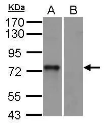
SYK antibody [N2C2], Internal detects SYK protein by western blot analysis. A. 30 μg mouse BMDM (bone marrow-derived macrophage) cells B. 30 μg mouse Syk null cells 10% SDS-PAGE SYK antibody [N2C2], Internal (GTX100748) dilution: 1:1000 The HRP-conjugated anti-rabbit IgG antibody (GTX213110-01) was used to detect the primary antibody.
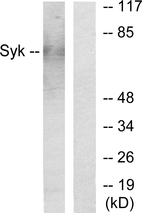
 was separated by 7.5% SDS-PAGE, and the membrane was blotted with SYK antibody (GTX100748) at a dilution of 1:2000. The HRP-conjugated anti-rabbit IgG antibody (GTX213110-01) was used to detect the primary antibody.")
 antibody at 1:200 dilution.")
![Immunoprecipitation of SYK protein from A431 whole cell extracts using 5 μg of SYK antibody [N2C2] (GTX100748). Western blot analysis was performed using SYK antibody [N2C2] (GTX100748). EasyBlot anti-Rabbit IgG (GTX221666-01) was used as a secondary reagent.](https://www.genetex.com/upload/website/prouct_img/normal/GTX100748/GTX100748_41892_20150112_IP_w_23060100_937.webp "Immunoprecipitation of SYK protein from A431 whole cell extracts using 5 μg of SYK antibody [N2C2] (GTX100748). Western blot analysis was performed using SYK antibody [N2C2] (GTX100748). EasyBlot anti-Rabbit IgG (GTX221666-01) was used as a secondary reagent.")
![SYK antibody [N2C2], Internal detects SYK protein at cytoplasm in human endometrium by immunohistochemical analysis. Sample: Paraffin-embedded human endometrium. SYK antibody [N2C2], Internal (GTX100748) diluted at 1:500.
Antigen Retrieval: Citrate buffer, pH 6.0, 15 min](https://www.genetex.com/upload/website/prouct_img/normal/GTX100748/GTX100748_41990_20150518_IHC-P_2_w_23060100_482.webp "SYK antibody [N2C2], Internal detects SYK protein at cytoplasm in human endometrium by immunohistochemical analysis. Sample: Paraffin-embedded human endometrium. SYK antibody [N2C2], Internal (GTX100748) diluted at 1:500.
Antigen Retrieval: Citrate buffer, pH 6.0, 15 min")
![SYK antibody [N2C2], Internal detects SYK protein at cytosol on rat spleen by immunohistochemical analysis. Sample: Paraffin-embedded rat spleen. SYK antibody [N2C2], Internal (GTX100748) dilution: 1:500.
Antigen Retrieval: Trilogy? (EDTA based, pH 8.0) buffer, 15min](https://www.genetex.com/upload/website/prouct_img/normal/GTX100748/GTX100748_40051_20141024_IHC_R_w_23060100_182.webp "SYK antibody [N2C2], Internal detects SYK protein at cytosol on rat spleen by immunohistochemical analysis. Sample: Paraffin-embedded rat spleen. SYK antibody [N2C2], Internal (GTX100748) dilution: 1:500.
Antigen Retrieval: Trilogy? (EDTA based, pH 8.0) buffer, 15min")
 was separated by 7.5% SDS-PAGE, and the membrane was blotted with SYK antibody (GTX100748) diluted by 1:1000. The HRP-conjugated anti-rabbit IgG antibody (GTX213110-01) was used to detect the primary antibody.")
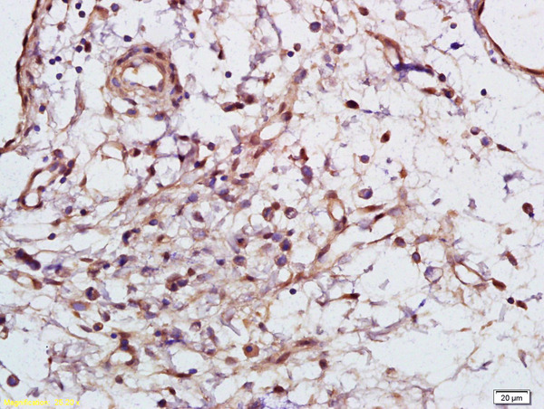
![SYK antibody [N2C2], Internal detects SYK protein at cytoplasm in human cervical cancer by immunohistochemical analysis. Sample: Paraffin-embedded human cervical cancer. SYK antibody [N2C2], Internal (GTX100748) diluted at 1:500.
Antigen Retrieval: Citrate buffer, pH 6.0, 15 min](https://www.genetex.com/upload/website/prouct_img/normal/GTX100748/GTX100748_41990_20150518_IHC-P_w_23060100_135.webp "SYK antibody [N2C2], Internal detects SYK protein at cytoplasm in human cervical cancer by immunohistochemical analysis. Sample: Paraffin-embedded human cervical cancer. SYK antibody [N2C2], Internal (GTX100748) diluted at 1:500.
Antigen Retrieval: Citrate buffer, pH 6.0, 15 min")
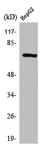
![Whole cell extract (30 μg) was separated by 7.5% SDS-PAGE, and the membrane was blotted with Syk antibody [N2C2], Internal (GTX100748) diluted at 1:1000. The HRP-conjugated anti-rabbit IgG antibody (GTX213110-01) was used to detect the primary antibody.](https://www.genetex.com/upload/website/prouct_img/normal/GTX100748/GTX100748_41906_20231222_WB_23122619_225.webp "Whole cell extract (30 μg) was separated by 7.5% SDS-PAGE, and the membrane was blotted with Syk antibody [N2C2], Internal (GTX100748) diluted at 1:1000. The HRP-conjugated anti-rabbit IgG antibody (GTX213110-01) was used to detect the primary antibody.")
SYK antibody [N2C2], Internal detects SYK protein by western blot analysis. A. 30 μg mouse BMDM (bone marrow-derived macrophage) cells B. 30 μg mouse Syk null cells 10% SDS-PAGE SYK antibody [N2C2], Internal (GTX100748) dilution: 1:1000 The HRP-conjugated anti-rabbit IgG antibody (GTX213110-01) was used to detect the primary antibody.
Syk antibody [N2C2], Internal
GTX100748
ApplicationsImmunoFluorescence, ImmunoPrecipitation, Western Blot, ImmunoCytoChemistry, ImmunoHistoChemistry, ImmunoHistoChemistry Paraffin
Product group Antibodies
ReactivityHuman, Mouse, Rat
TargetSYK
Overview
- SupplierGeneTex
- Product NameSyk antibody [N2C2], Internal
- Delivery Days Customer9
- Application Supplier NoteWB: 1:500-1:3000. ICC/IF: 1:100-1:1000. IHC-P: 1:100-1:1000. IP: 1:100-1:500. *Optimal dilutions/concentrations should be determined by the researcher.Not tested in other applications.
- ApplicationsImmunoFluorescence, ImmunoPrecipitation, Western Blot, ImmunoCytoChemistry, ImmunoHistoChemistry, ImmunoHistoChemistry Paraffin
- CertificationResearch Use Only
- ClonalityPolyclonal
- Concentration0.32 mg/ml
- ConjugateUnconjugated
- Gene ID6850
- Target nameSYK
- Target descriptionspleen associated tyrosine kinase
- Target synonymsIMD82, p72-Syk, tyrosine-protein kinase SYK, spleen tyrosine kinase
- HostRabbit
- IsotypeIgG
- Protein IDP43405
- Protein NameTyrosine-protein kinase SYK
- Scientific DescriptionThis gene encodes a member of the family of non-receptor type Tyr protein kinases. This protein is widely expressed in hematopoietic cells and is involved in coupling activated immunoreceptors to downstream signaling events that mediate diverse cellular responses, including proliferation, differentiation, and phagocytosis. It is thought to be a modulator of epithelial cell growth and a potential tumour suppressor in human breast carcinomas. Alternatively spliced transcript variants encoding different isoforms have been found for this gene. [provided by RefSeq]
- ReactivityHuman, Mouse, Rat
- Storage Instruction-20°C or -80°C,2°C to 8°C
- UNSPSC41116161
Datasheet
Related products
Product group Antibodies

Anti-pSYK [MIL81-1-8]Ab02455-1.1
ApplicationsImmunoFluorescence, ELISA, ImmunoCytoChemistry, ImmunoHistoChemistry
ReactivityHuman
TargetSYK
- SizePrice
Product group Antibodies
Anti-SYK AntibodyA101587
ApplicationsWestern Blot, ELISA
ReactivityHuman
- SizePrice
Product group Antibodies
Anti-SYK Antibody144-02123
ApplicationsImmunoFluorescence, Western Blot, ImmunoHistoChemistry
ReactivityHuman, Mouse, Rat
TargetSYK
- SizePrice
Product group Antibodies

SYK AntibodyLS-C761163
ApplicationsImmunoFluorescence, Western Blot, ImmunoCytoChemistry, ImmunoHistoChemistry
ReactivityHuman, Mouse, Rat
TargetSYK
- SizePrice

Product group Antibodies
Anti-SYK Antibody Picoband(r)A00490-3-CARRIER-FREE
ApplicationsFlow Cytometry, Western Blot, ELISA
ReactivityHuman, Mouse, Rat
TargetSYK
- SizePrice
Product group Antibodies
References
ApplicationsImmunoFluorescence, Western Blot, ELISA, ImmunoCytoChemistry, ImmunoHistoChemistry, ImmunoHistoChemistry Frozen, ImmunoHistoChemistry Paraffin
ReactivityBovine, Canine, Chicken, Guinea Pig, Human, Mouse, Porcine, Rabbit, Rat
TargetSYK
- SizePrice
Product group Antibodies
SYK AntibodyCSB-PA004198
ApplicationsWestern Blot, ELISA, ImmunoHistoChemistry
ReactivityHuman
TargetSYK
- SizePrice
Product group Antibodies

ApplicationsWestern Blot, ELISA
ReactivityBovine, Canine, Human, Mouse, Porcine, Rat
TargetSYK
- SizePrice
Product group Antibodies
ApplicationsWestern Blot, ImmunoHistoChemistry
ReactivityMouse, Porcine, Rat
TargetSYK
- SizePrice