

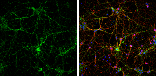
Synaptophysin antibody detects Synaptophysin protein at synaptic vesicles by immunofluorescent analysis. Sample: DIV9 rat E18 primary cortical neurons were fixed in 4% paraformaldehyde at RT for 15 min. Green: Synaptophysin protein stained by Synaptophysin antibody (GTX100865) diluted at 1:500. Red: beta Tubulin 3/ Tuj1, stained by beta Tubulin 3/ Tuj1 antibody [GT11710] (GTX631836) diluted at 1:500. Blue: Fluoroshield with DAPI (GTX30920).
![Synaptophysin antibody detects Synaptophysin protein by immunofluorescent analysis. Sample: DIV10 rat E18 primary hippocampal neuron cells were fixed in 4% paraformaldehyde at RT for 15 min. Green: Synaptophysin stained by Synaptophysin antibody (GTX100865) diluted at 1:500. Red: Tau, stained by Tau antibody [GT287] (GTX634809) diluted at 1:500. Blue: Fluoroshield with DAPI (GTX30920).](https://www.genetex.com/upload/website/prouct_img/normal/GTX100865/GTX100865_43733_20191125_ICC_IF_R_w_23060100_123.webp "Synaptophysin antibody detects Synaptophysin protein by immunofluorescent analysis. Sample: DIV10 rat E18 primary hippocampal neuron cells were fixed in 4% paraformaldehyde at RT for 15 min. Green: Synaptophysin stained by Synaptophysin antibody (GTX100865) diluted at 1:500. Red: Tau, stained by Tau antibody [GT287] (GTX634809) diluted at 1:500. Blue: Fluoroshield with DAPI (GTX30920).")
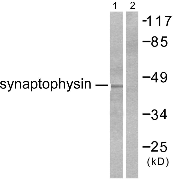
 were separated by 12% SDS-PAGE, and the membrane was blotted with Synaptophysin antibody (GTX100865) diluted at 1:50000. The HRP-conjugated anti-rabbit IgG antibody (GTX213110-01) was used to detect the primary antibody.")
 dilution: 1:500.
Antigen Retrieval: Trilogy? (EDTA based, pH 8.0) buffer, 15min")
 diluted at 1:500. Antigen Retrieval: Citrate buffer, pH 6.0, 15 min")
, p63 (GTX102425) and Cytokeratin 7 (GTX109723) in human small cell lung cancer (SCLC) and non-small cell lung cancer (NSCLC) specimens. Sample: Paraffin-embedded human SCLC (upper panel) and NSCLC (lower panel). The section was pre-treated using heat mediated antigen retrieval with sodium citrate buffer (pH6) for 15 mins. The section was then incubated with primary antibody at 1:500 overnight at 4oC and detected using an HRP conjugated avidin-biotin-peroxidase Complex system. DAB was used as the chromogen and counterstained with haematoxylin.
Antigen Retrieval: Citrate buffer, pH 6.0, 15 min")
 diluted at 1:250. Red: α-Bungarotoxin, stained by α-Bungarotoxin, Alexa Fluor? 594 conjugate (B13423) diluted at 1:5000. Blue: Hoechst 33342 staining.")
 was separated by 12% SDS-PAGE, and the membrane was blotted with Synaptophysin antibody (GTX100865) diluted at 1:50000. The HRP-conjugated anti-rabbit IgG antibody (GTX213110-01) was used to detect the primary antibody.")
 antibody at 1:100 dilution.
Antigen Retrieval: Trilogy? (EDTA based, pH 8.0) buffer, 15min")
![Synaptophysin antibody detects Synaptophysin protein expression by immunohistochemical analysis. Sample: Paraffin-Embedded adult mouse retina. Green: Synaptophysin protein stained by Synaptophysin antibody (GTX100865) diluted at 1:250. Red: beta Tubulin 3/ TUJ1, stained by beta Tubulin 3/ TUJ1 antibody [GT11710] (GTX631836) diluted at 1:250. Blue: Fluoroshield with DAPI (GTX30920).
Antigen Retrieval: Citrate buffer, pH 6.0, 15 min](https://www.genetex.com/upload/website/prouct_img/normal/GTX100865/GTX100865_42347_20170306_IHC-P_M_w_23060100_512.webp "Synaptophysin antibody detects Synaptophysin protein expression by immunohistochemical analysis. Sample: Paraffin-Embedded adult mouse retina. Green: Synaptophysin protein stained by Synaptophysin antibody (GTX100865) diluted at 1:250. Red: beta Tubulin 3/ TUJ1, stained by beta Tubulin 3/ TUJ1 antibody [GT11710] (GTX631836) diluted at 1:250. Blue: Fluoroshield with DAPI (GTX30920).
Antigen Retrieval: Citrate buffer, pH 6.0, 15 min")
Synaptophysin antibody detects Synaptophysin protein at synaptic vesicles by immunofluorescent analysis. Sample: DIV9 rat E18 primary cortical neurons were fixed in 4% paraformaldehyde at RT for 15 min. Green: Synaptophysin protein stained by Synaptophysin antibody (GTX100865) diluted at 1:500. Red: beta Tubulin 3/ Tuj1, stained by beta Tubulin 3/ Tuj1 antibody [GT11710] (GTX631836) diluted at 1:500. Blue: Fluoroshield with DAPI (GTX30920).
Synaptophysin antibody
GTX100865
ApplicationsImmunoFluorescence, Western Blot, ImmunoCytoChemistry, ImmunoHistoChemistry, ImmunoHistoChemistry Frozen, ImmunoHistoChemistry Paraffin
Product group Antibodies
ReactivityHuman, Mouse, Rat
TargetSYP
Overview
- SupplierGeneTex
- Product NameSynaptophysin antibody
- Delivery Days Customer9
- Application Supplier NoteWB: 1:500-1:50000. ICC/IF: 1:100-1:1000. IHC-P: 1:100-1:1000. *Optimal dilutions/concentrations should be determined by the researcher.Not tested in other applications.
- ApplicationsImmunoFluorescence, Western Blot, ImmunoCytoChemistry, ImmunoHistoChemistry, ImmunoHistoChemistry Frozen, ImmunoHistoChemistry Paraffin
- CertificationResearch Use Only
- ClonalityPolyclonal
- Concentration0.65 mg/ml
- ConjugateUnconjugated
- Gene ID6855
- Target nameSYP
- Target descriptionsynaptophysin
- Target synonymsMRX96, MRXSYP, XLID96, synaptophysin, major synaptic vesicle protein P38
- HostRabbit
- IsotypeIgG
- Protein IDP08247
- Protein NameSynaptophysin
- Scientific DescriptionSynaptophysin (p38) is an integral membrane protein of small synaptic vesicles in brain and endocrine cells.[supplied by OMIM]
- ReactivityHuman, Mouse, Rat
- Storage Instruction-20°C or -80°C,2°C to 8°C
- UNSPSC41116161
Datasheet
Related products
Product group Antibodies
ApplicationsWestern Blot, ELISA, ImmunoHistoChemistry
ReactivityHuman, Mouse, Rat
- SizePrice

Product group Antibodies
Anti-Synaptophysin/SYP Picoband(r) AntibodyA05049-CARRIER-FREE
ApplicationsImmunoFluorescence, Western Blot, ImmunoCytoChemistry, ImmunoHistoChemistry
ReactivityHuman, Mouse, Rat
TargetSYP
- SizePrice
Product group Antibodies

Synaptophysin (SYP) AntibodyABX013199
ApplicationsWestern Blot, ELISA, ImmunoHistoChemistry
- SizePrice
Product group Antibodies
Anti-SYP Antibody144-06344
ApplicationsImmunoFluorescence, Western Blot, ImmunoHistoChemistry
ReactivityHuman, Mouse, Rat
TargetSYP
- SizePrice
Product group Antibodies

ApplicationsImmunoHistoChemistry
ReactivityHuman
TargetSYP
- SizePrice

Product group Antibodies
Synaptophysin Recombinant AntibodyBSM-52379R
ApplicationsFlow Cytometry, ImmunoFluorescence, Western Blot, ImmunoHistoChemistry, ImmunoHistoChemistry Frozen, ImmunoHistoChemistry Paraffin
ReactivityHuman, Mouse, Rat
TargetSYP
- SizePrice
Product group Antibodies
SYP AntibodyCSB-PA004215
ApplicationsWestern Blot, ELISA, ImmunoHistoChemistry
ReactivityHuman, Mouse, Rat
TargetSYP
- SizePrice
Product group Antibodies
ApplicationsImmunoPrecipitation, Western Blot, ImmunoCytoChemistry, ImmunoHistoChemistry
TargetSYP
- SizePrice
Product group Antibodies
Synaptophysin antibodyGTX14692
ApplicationsImmunoFluorescence, ImmunoPrecipitation, Western Blot, ELISA, ImmunoCytoChemistry, ImmunoHistoChemistry, ImmunoHistoChemistry Paraffin
ReactivityChicken, Human, Monkey, Mouse, Rat
TargetSYP
- SizePrice
![IHC-P analysis of human pancreas tissue using GTX26245 Synaptophysin antibody [SYP02].](https://www.genetex.com/upload/website/prouct_img/normal/GTX26245/GTX26245_20191203_IHC-P_114_w_23060722_767.webp)
Product group Antibodies
Synaptophysin antibody [SYP02]GTX26245
ApplicationsImmunoHistoChemistry, ImmunoHistoChemistry Paraffin
ReactivityHuman
TargetSYP
- SizePrice