

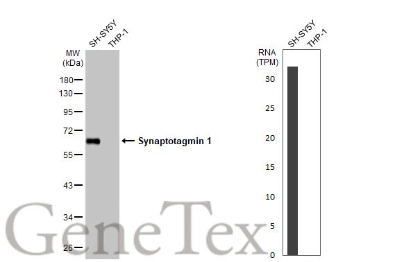
Various whole cell extracts (30 μg) were separated by 10% SDS-PAGE, and the membrane was blotted with Synaptotagmin 1 antibody [HL1626] (GTX637119) diluted at 1:2000. The HRP-conjugated anti-rabbit IgG antibody (GTX213110-01) was used to detect the primary antibody, and the signal was developed with Trident ECL plus-Enhanced. Corresponding RNA expression data for the same cell lines are based on Human Protein Atlas program.
![Human tissue extract (5 μg) was separated by 10% SDS-PAGE, and the membrane was blotted with Synaptotagmin 1 antibody [HL1626] (GTX637119) diluted at 1:20000. The HRP-conjugated anti-rabbit IgG antibody (GTX213110-01) was used to detect the primary antibody.](https://www.genetex.com/upload/website/prouct_img/normal/GTX637119/GTX637119_T-44739_20221111_WB_brain_22111518_711.webp "Human tissue extract (5 μg) was separated by 10% SDS-PAGE, and the membrane was blotted with Synaptotagmin 1 antibody [HL1626] (GTX637119) diluted at 1:20000. The HRP-conjugated anti-rabbit IgG antibody (GTX213110-01) was used to detect the primary antibody.")
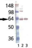
![Various tissue extracts (50 μg) were separated by 10% SDS-PAGE, and the membrane was blotted with Synaptotagmin 1 antibody [HL1626] (GTX637119) diluted at 1:20000. The HRP-conjugated anti-rabbit IgG antibody (GTX213110-01) was used to detect the primary antibody.](https://www.genetex.com/upload/website/prouct_img/normal/GTX637119/GTX637119_T-44739_20221111_WB_M_R_22111518_736.webp "Various tissue extracts (50 μg) were separated by 10% SDS-PAGE, and the membrane was blotted with Synaptotagmin 1 antibody [HL1626] (GTX637119) diluted at 1:20000. The HRP-conjugated anti-rabbit IgG antibody (GTX213110-01) was used to detect the primary antibody.")
![Drosophila tissue extract (50 μg) was separated by 10% SDS-PAGE, and the membrane was blotted with Synaptotagmin 1 antibody [HL1626] (GTX637119) diluted at 1:1000. The HRP-conjugated anti-rabbit IgG antibody (GTX213110-01) was used to detect the primary antibody.](https://www.genetex.com/upload/website/prouct_img/normal/GTX637119/GTX637119_44886_20230324_WB_Drosophila_brain_23032819_220.webp "Drosophila tissue extract (50 μg) was separated by 10% SDS-PAGE, and the membrane was blotted with Synaptotagmin 1 antibody [HL1626] (GTX637119) diluted at 1:1000. The HRP-conjugated anti-rabbit IgG antibody (GTX213110-01) was used to detect the primary antibody.")
![Synaptotagmin 1 antibody [HL1626] detects Synaptotagmin 1 protein at cell membrane and cytoplasm by immunohistochemical analysis. Sample: Paraffin-embedded rat eye. Synaptotagmin 1 stained by Synaptotagmin 1 antibody [HL1626] (GTX637119) diluted at 1:250. Red: beta Tubulin 3/ Tuj1, a neural marker, stained by beta Tubulin 3/ Tuj1 antibody [GT11710] (GTX631836) diluted at 1:500. Blue: Fluoroshield with DAPI (GTX30920). Antigen Retrieval: Citrate buffer, pH 6.0, 15 min](https://www.genetex.com/upload/website/prouct_img/normal/GTX637119/GTX637119_T-44739_20240401_IHC-P_R_24041019_311.webp "Synaptotagmin 1 antibody [HL1626] detects Synaptotagmin 1 protein at cell membrane and cytoplasm by immunohistochemical analysis. Sample: Paraffin-embedded rat eye. Synaptotagmin 1 stained by Synaptotagmin 1 antibody [HL1626] (GTX637119) diluted at 1:250. Red: beta Tubulin 3/ Tuj1, a neural marker, stained by beta Tubulin 3/ Tuj1 antibody [GT11710] (GTX631836) diluted at 1:500. Blue: Fluoroshield with DAPI (GTX30920). Antigen Retrieval: Citrate buffer, pH 6.0, 15 min")
Various whole cell extracts (30 μg) were separated by 10% SDS-PAGE, and the membrane was blotted with Synaptotagmin 1 antibody [HL1626] (GTX637119) diluted at 1:2000. The HRP-conjugated anti-rabbit IgG antibody (GTX213110-01) was used to detect the primary antibody, and the signal was developed with Trident ECL plus-Enhanced. Corresponding RNA expression data for the same cell lines are based on Human Protein Atlas program.
Synaptotagmin 1 antibody [HL1626]
GTX637119
ApplicationsWestern Blot, ImmunoHistoChemistry, ImmunoHistoChemistry Paraffin
Product group Antibodies
ReactivityDrosophila, Human, Mouse, Rat
TargetSYT1
Overview
- SupplierGeneTex
- Product NameSynaptotagmin 1 antibody [HL1626]
- Delivery Days Customer12
- ApplicationsWestern Blot, ImmunoHistoChemistry, ImmunoHistoChemistry Paraffin
- CertificationResearch Use Only
- ClonalityMonoclonal
- Clone IDHL1626
- Concentration1 mg/ml
- ConjugateUnconjugated
- Gene ID6857
- Target nameSYT1
- Target descriptionsynaptotagmin 1
- Target synonymsBAGOS, P65, SVP65, SYT, synaptotagmin-1, synaptotagmin I, sytI
- HostRabbit
- IsotypeIgG
- Protein IDP21579
- Protein NameSynaptotagmin-1
- Scientific DescriptionThe synaptotagmins are integral membrane proteins of synaptic vesicles thought to serve as Ca(2+) sensors in the process of vesicular trafficking and exocytosis. Calcium binding to synaptotagmin-1 participates in triggering neurotransmitter release at the synapse (Fernandez-Chacon et al., 2001 [PubMed 11242035]).[supplied by OMIM, Jul 2010]
- ReactivityDrosophila, Human, Mouse, Rat
- Storage Instruction-20°C or -80°C,2°C to 8°C
- UNSPSC41116161
Datasheet
Related products
Product group Antibodies
ApplicationsWestern Blot, ELISA, ImmunoHistoChemistry
ReactivityHuman, Mouse, Rat
- SizePrice
Product group Antibodies

ApplicationsWestern Blot, ELISA, ImmunoHistoChemistry
- SizePrice
Product group Antibodies
Anti-SYT1 Antibody144-61669
ApplicationsImmunoFluorescence, Western Blot, ImmunoHistoChemistry
ReactivityHuman, Mouse, Rat
TargetSYT1
- SizePrice

Product group Antibodies
Anti-Synaptotagmin 1/SYT1 Antibody Picoband(r)A02314-1-CARRIER-FREE
ApplicationsFlow Cytometry, Western Blot, ImmunoCytoChemistry, ImmunoHistoChemistry
ReactivityHuman, Mouse, Rat
TargetSYT1
- SizePrice
Product group Antibodies
ApplicationsImmunoFluorescence, Western Blot, ELISA, ImmunoCytoChemistry, ImmunoHistoChemistry, ImmunoHistoChemistry Frozen, ImmunoHistoChemistry Paraffin
ReactivityBovine, Canine, Chicken, Human, Mouse, Porcine, Rabbit, Rat
TargetSYT1
- SizePrice
Product group Antibodies
SYT1/SYT2 AntibodyCSB-PA004203
ApplicationsWestern Blot, ELISA, ImmunoHistoChemistry
ReactivityHuman, Mouse, Rat
TargetSYT1
- SizePrice
Product group Antibodies

Goat anti-Synaptotagmin IEB08253
ApplicationsWestern Blot, ELISA, ImmunoHistoChemistry
ReactivityBovine, Canine, Human, Mouse, Rat
TargetSYT1
- SizePrice
Product group Antibodies
ApplicationsImmunoPrecipitation, Western Blot, ImmunoCytoChemistry, ImmunoHistoChemistry
ReactivityMouse, Porcine, Rat
TargetSYT1
- SizePrice
Product group Antibodies
Synaptotagmin antibody [ASV30]GTX13259
ApplicationsImmunoFluorescence, ImmunoPrecipitation, Western Blot, ImmunoCytoChemistry, ImmunoHistoChemistry
ReactivityBovine, Fish, Mouse, Rabbit, Rat, Xenopus
TargetSYT1
- SizePrice
Product group Antibodies

SYT1 / Synaptotagmin Antibody (Internal)LS-C358873
ApplicationsImmunoFluorescence, Western Blot, ImmunoCytoChemistry, ImmunoHistoChemistry, ImmunoHistoChemistry Paraffin
ReactivityHuman, Mouse, Rat
TargetSYT1
- SizePrice