

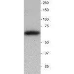
WB analysis of neonatal mouse brain homogenate using GTX85467 Synaptotagmin antibody. Loading : 25 ug


WB analysis of neonatal mouse brain homogenate using GTX85467 Synaptotagmin antibody. Loading : 25 ug
Synaptotagmin antibody
GTX85467
ApplicationsImmunoFluorescence, Western Blot, ImmunoCytoChemistry, ImmunoHistoChemistry, ImmunoHistoChemistry Frozen
Product group Antibodies
ReactivityHuman, Mouse, Rat
TargetSYT1
Overview
- SupplierGeneTex
- Product NameSynaptotagmin antibody
- Delivery Days Customer9
- Application Supplier NoteWB: 1:1000-1:2000. IHC: 1:1000-1:2000. *Optimal dilutions/concentrations should be determined by the researcher.Not tested in other applications.
- ApplicationsImmunoFluorescence, Western Blot, ImmunoCytoChemistry, ImmunoHistoChemistry, ImmunoHistoChemistry Frozen
- CertificationResearch Use Only
- ClonalityPolyclonal
- Concentration400 ug/ml
- ConjugateUnconjugated
- Gene ID6857
- Target nameSYT1
- Target descriptionsynaptotagmin 1
- Target synonymsBAGOS, P65, SVP65, SYT, synaptotagmin-1, synaptotagmin I, sytI
- HostChicken
- IsotypeIgY
- Scientific DescriptionHuman Synaptotagmin-1 is a 47,442 dalton protein (422 amino acids) associated with pre-synaptic terminals of the PNS and CNS. Synaptotagmin-1 binds calcium in synaptic terminals and initiates the fusion of vesicles with the pre-synaptic membrane. This protein is also believed to be the target of proteolytic activity associated with Botulinum neurotoxin A.
- ReactivityHuman, Mouse, Rat
- Storage Instruction2°C to 8°C
- UNSPSC12352203
Datasheet
Related products
Product group Antibodies

ApplicationsWestern Blot, ELISA, ImmunoHistoChemistry
- SizePrice
Product group Antibodies
Anti-SYT1 Antibody144-61669
ApplicationsImmunoFluorescence, Western Blot, ImmunoHistoChemistry
ReactivityHuman, Mouse, Rat
TargetSYT1
- SizePrice

Product group Antibodies
Anti-Synaptotagmin 1/SYT1 Antibody Picoband(r)A02314-1-CARRIER-FREE
ApplicationsFlow Cytometry, Western Blot, ImmunoCytoChemistry, ImmunoHistoChemistry
ReactivityHuman, Mouse, Rat
TargetSYT1
- SizePrice
![Various whole cell extracts (30 Aμg) were separated by 10% SDS-PAGE, and the membrane was blotted with Synaptotagmin 1 antibody [N1C2] (GTX114124) diluted at 1:10000. The HRP-conjugated anti-rabbit IgG antibody (GTX213110-01) was used to detect the primary antibody, and the signal was developed with Trident ECL plus-Enhanced. Corresponding RNA expression data for the same cell lines are based on Human Protein Atlas program.](https://www.genetex.com/upload/website/prouct_img/normal/GTX114124/GTX114124_40583_20220805_WB_TPM_watermark_22080923_835.webp)
Product group Antibodies
Synaptotagmin 1 antibody [N1C2]GTX114124
ApplicationsImmunoFluorescence, Western Blot, ImmunoCytoChemistry, ImmunoHistoChemistry, ImmunoHistoChemistry Paraffin
ReactivityHuman, Mouse, Rat
TargetSYT1
- SizePrice
![Various whole cell extracts (30 μg) were separated by 10% SDS-PAGE, and the membrane was blotted with Synaptotagmin 1 antibody [HL1626] (GTX637119) diluted at 1:2000. The HRP-conjugated anti-rabbit IgG antibody (GTX213110-01) was used to detect the primary antibody, and the signal was developed with Trident ECL plus-Enhanced. Corresponding RNA expression data for the same cell lines are based on Human Protein Atlas program.](https://www.genetex.com/upload/website/prouct_img/normal/GTX637119/GTX637119_T-44739_20221021_WB_TPM_watermark_22102723_356.webp)
Product group Antibodies
Synaptotagmin 1 antibody [HL1626]GTX637119
ApplicationsWestern Blot, ImmunoHistoChemistry, ImmunoHistoChemistry Paraffin
ReactivityDrosophila, Human, Mouse, Rat
TargetSYT1
- SizePrice
![Various tissue extracts (50 μg) were separated by 10% SDS-PAGE, and the membrane was blotted with Synaptotagmin 1 antibody [HL1654] (GTX637252) diluted at 1:20000. The HRP-conjugated anti-rabbit IgG antibody (GTX213110-01) was used to detect the primary antibody.](https://www.genetex.com/upload/website/prouct_img/normal/GTX637252/GTX637252_T-44753_20221111_WB_M_R_22111518_179.webp)
Product group Antibodies
Synaptotagmin 1 antibody [HL1654]GTX637252
ApplicationsWestern Blot, ImmunoHistoChemistry, ImmunoHistoChemistry Paraffin
ReactivityDrosophila, Human, Mouse, Rat
TargetSYT1
- SizePrice
Product group Antibodies
Synaptotagmin I/II antibodyGTX78914
ApplicationsWestern Blot
ReactivityHuman
TargetSYT1
- SizePrice
Product group Antibodies
ApplicationsImmunoPrecipitation, Western Blot, ImmunoCytoChemistry, ImmunoHistoChemistry
ReactivityMouse, Porcine, Rat
TargetSYT1
- SizePrice
Product group Antibodies
ApplicationsWestern Blot, ImmunoHistoChemistry, ImmunoHistoChemistry Paraffin
ReactivityHuman
TargetSYT1
- SizePrice