

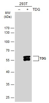
TDG antibody detects TDG protein by western blot analysis. 293T mock transfected (-) or transfected with a construct expressing human TDG. 10 % SDS-PAGE TDG antibody (GTX631589) dilution: 1:10000
TDG antibody detects TDG protein by western blot analysis. 293T mock transfected (-) or transfected with a construct expressing human TDG. 10 % SDS-PAGE TDG antibody (GTX631589) dilution: 1:10000
TDG antibody [GT622]
GTX631589
ApplicationsWestern Blot
Product group Antibodies
ReactivityHuman
TargetTDG
Overview
- SupplierGeneTex
- Product NameTDG antibody [GT622]
- Delivery Days Customer9
- Application Supplier NoteWB: 1:5000-1:20000. *Optimal dilutions/concentrations should be determined by the researcher.Not tested in other applications.
- ApplicationsWestern Blot
- CertificationResearch Use Only
- ClonalityMonoclonal
- Clone IDGT622
- Concentration1 mg/ml
- ConjugateUnconjugated
- Gene ID6996
- Target nameTDG
- Target descriptionthymine DNA glycosylase
- Target synonymshTDG, G/T mismatch-specific thymine DNA glycosylase
- HostMouse
- IsotypeIgG1
- Protein IDQ13569
- Protein NameG/T mismatch-specific thymine DNA glycosylase
- Scientific DescriptionThe protein encoded by this gene belongs to the TDG/mug DNA glycosylase family. Thymine-DNA glycosylase (TDG) removes thymine moieties from G/T mismatches by hydrolyzing the carbon-nitrogen bond between the sugar-phosphate backbone of DNA and the mispaired thymine. With lower activity, this enzyme also removes thymine from C/T and T/T mispairings. TDG can also remove uracil and 5-bromouracil from mispairings with guanine. This enzyme plays a central role in cellular defense against genetic mutation caused by the spontaneous deamination of 5-methylcytosine and cytosine. This gene may have a pseudogene in the p arm of chromosome 12. [provided by RefSeq]
- ReactivityHuman
- Storage Instruction-20°C or -80°C,2°C to 8°C
- UNSPSC41116161
Datasheet
Related products

Product group Antibodies
Anti-TDG Antibody Picoband(r)A15317-1-CARRIER-FREE
ApplicationsFlow Cytometry, Western Blot, ELISA
ReactivityHuman, Monkey
TargetTDG
- SizePrice
Product group Antibodies
Anti-TDG Antibody144-05756
ApplicationsWestern Blot
ReactivityHuman, Mouse, Rat
TargetTDG
- SizePrice
Product group Antibodies
Anti-TDG AntibodyA98428
ApplicationsWestern Blot, ELISA
ReactivityHuman, Mouse, Rat
- SizePrice
Product group Antibodies

TDG / Thymine DNA Glycosylase AntibodyLS-C748418
ApplicationsImmunoFluorescence, Western Blot
ReactivityHuman, Mouse, Rat
TargetTDG
- SizePrice
Product group Antibodies
TDG AntibodyCSB-PA050229
ApplicationsImmunoFluorescence, Western Blot, ELISA, ImmunoHistoChemistry
ReactivityHuman, Mouse, Rat
TargetTDG
- SizePrice

Product group Antibodies
Anti-TDG AntibodyHPA052263
ApplicationsImmunoCytoChemistry, ImmunoHistoChemistry
ReactivityHuman
TargetTDG
- SizePrice

Product group Antibodies
TDG antibodyGTX110473
ApplicationsImmunoFluorescence, Western Blot, ImmunoCytoChemistry
ReactivityHuman, Mouse
TargetTDG
- SizePrice
Product group Antibodies
TDG antibodyGTX127975
ApplicationsWestern Blot
ReactivityHuman, Rat
TargetTDG
- SizePrice
Product group Antibodies
TDG antibodyGTX129486
ApplicationsWestern Blot
ReactivityHuman
TargetTDG
- SizePrice