

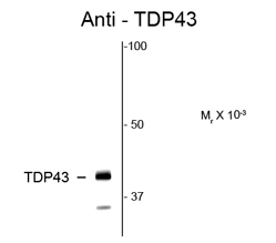
Western blot of rat hippocampal lysate showing specific immunolabeling of the ~58k TR-α2 protein using TDP43 antibody (GTX82580).
Western blot of rat hippocampal lysate showing specific immunolabeling of the ~58k TR-α2 protein using TDP43 antibody (GTX82580).
TDP43 antibody
GTX82580
ApplicationsWestern Blot, ImmunoHistoChemistry
Product group Antibodies
ReactivityHuman, Rat
TargetTARDBP
Overview
- SupplierGeneTex
- Product NameTDP43 antibody
- Delivery Days Customer9
- ApplicationsWestern Blot, ImmunoHistoChemistry
- CertificationResearch Use Only
- ClonalityPolyclonal
- ConjugateUnconjugated
- Gene ID23435
- Target nameTARDBP
- Target descriptionTAR DNA binding protein
- Target synonymsALS10, TDP-43, TAR DNA-binding protein 43, TAR DNA-binding protein-43
- HostRabbit
- IsotypeIgG
- Protein IDQ13148
- Protein NameTAR DNA-binding protein 43
- Scientific DescriptionHIV-1, the causative agent of acquired immunodeficiency syndrome (AIDS), contains an RNA genome that produces a chromosomally integrated DNA during the replicative cycle. Activation of HIV-1 gene expression by the transactivator Tat is dependent on an RNA regulatory element (TAR) located downstream of the transcription initiation site. The protein encoded by this gene is a transcriptional repressor that binds to chromosomally integrated TAR DNA and represses HIV-1 transcription. In addition, this protein regulates alternate splicing of the CFTR gene. A similar pseudogene is present on chromosome 20. [provided by RefSeq, Jul 2008]
- ReactivityHuman, Rat
- Storage Instruction-20°C or -80°C,2°C to 8°C
- UNSPSC41116161
References
- Neither a Novel Tau Proteinopathy nor an Expansion of a Phenotype: Reappraising Clinicopathology-Based Nosology. Marsili L et al., 2021 Jul 7, Int J Mol SciRead this paper
Datasheet
Related products
Product group Antibodies
TARDBP AntibodyCSB-PA023129EA01HU
ApplicationsImmunoFluorescence, Western Blot, ELISA, ImmunoHistoChemistry
ReactivityHuman
TargetTARDBP
- SizePrice

Product group Antibodies
Anti-TDP-43/TARDBP Antibody Picoband(r)A01001-3-CARRIER-FREE
ApplicationsFlow Cytometry, ImmunoFluorescence, Western Blot, ELISA, ImmunoCytoChemistry, ImmunoHistoChemistry
ReactivityHuman, Mouse, Rat
TargetTARDBP
- SizePrice
Product group Antibodies
Anti-TARDBP AntibodyA29471
ApplicationsImmunoFluorescence, ImmunoPrecipitation, Western Blot, ImmunoHistoChemistry, Other Application
ReactivityHuman, Mouse, Rat
- SizePrice
Product group Antibodies

TDP-43 / TARDBP AntibodyLS-C748455
ApplicationsImmunoFluorescence, Western Blot
ReactivityHuman, Mouse, Rat
TargetTARDBP
- SizePrice
Product group Antibodies

Goat anti-TDP-43EB09108
ApplicationsWestern Blot, ELISA, ImmunoHistoChemistry
ReactivityBovine, Canine, Human, Mouse, Porcine, Rat
TargetTARDBP
- SizePrice

Product group Antibodies
Anti-TARDBP AntibodyHPA017284
ApplicationsWestern Blot, ImmunoCytoChemistry, ImmunoHistoChemistry
ReactivityHuman
TargetTARDBP
- SizePrice
Product group Antibodies
References
TDP43 antibodyGTX30809
ApplicationsImmunoFluorescence, ImmunoPrecipitation, Western Blot, ELISA, ImmunoCytoChemistry, ImmunoHistoChemistry, ImmunoHistoChemistry Paraffin
ReactivityHuman, Mouse
TargetTARDBP
- SizePrice
Product group Antibodies
TARDBP Polyclonal AntibodyCAC14898
ApplicationsImmunoFluorescence, Western Blot, ELISA, ImmunoHistoChemistry
TargetTARDBP
- SizePrice
Product group Antibodies
TARDBP Recombinant AntibodyBSM-60837R
ApplicationsFlow Cytometry, ImmunoFluorescence, Western Blot, ImmunoCytoChemistry, ImmunoHistoChemistry, ImmunoHistoChemistry Frozen, ImmunoHistoChemistry Paraffin
ReactivityHuman, Mouse, Rat
TargetTARDBP
- SizePrice