

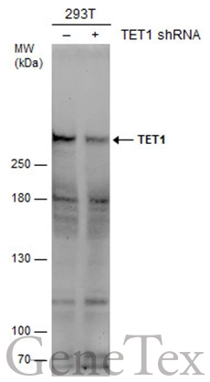
Non-transfected (–) and transfected (+) 293T whole cell extracts (50 μg) were separated by 5% SDS-PAGE, and the membrane was blotted with TET1 antibody [N3C1] (GTX124207) diluted at 1:1000. The HRP-conjugated anti-rabbit IgG antibody (GTX213110-01) was used to detect the primary antibody.
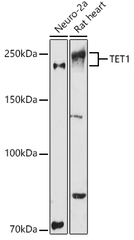
![Various whole cell extracts (30 μg) were separated by 5% SDS-PAGE, and the membrane was blotted with TET1 antibody [N3C1] (GTX124207) diluted at 1:2000. The HRP-conjugated anti-rabbit IgG antibody (GTX213110-01) was used to detect the primary antibody.](https://www.genetex.com/upload/website/prouct_img/normal/GTX124207/GTX124207_42915_20170714_WB_w_23060522_890.webp "Various whole cell extracts (30 μg) were separated by 5% SDS-PAGE, and the membrane was blotted with TET1 antibody [N3C1] (GTX124207) diluted at 1:2000. The HRP-conjugated anti-rabbit IgG antibody (GTX213110-01) was used to detect the primary antibody.")
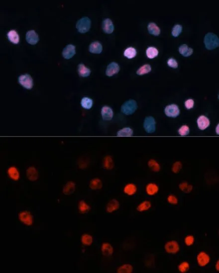
![TET1 antibody [N3C1] detects TET1 protein at nucleus by immunofluorescent analysis. Sample: Mock and transfected 293T cells were fixed in 4% paraformaldehyde at RT for 15 min. Green: TET1 stained by TET1 antibody [N3C1] (GTX124207) diluted at 1:1000. Blue: Hoechst 33342 staining.](https://www.genetex.com/upload/website/prouct_img/normal/GTX124207/GTX124207_42915_20180801_ICC_IF_B_w_23060522_953.webp "TET1 antibody [N3C1] detects TET1 protein at nucleus by immunofluorescent analysis. Sample: Mock and transfected 293T cells were fixed in 4% paraformaldehyde at RT for 15 min. Green: TET1 stained by TET1 antibody [N3C1] (GTX124207) diluted at 1:1000. Blue: Hoechst 33342 staining.")
 antibody at 1:1000 dilution.
Antigen Retrieval: Trilogy? (EDTA based, pH 8.0) buffer, 15min")
![TET1 antibody [N3C1] detects TET1 protein at cytoplasm and nucleus by immunofluorescent analysis. Sample: HeLa cells were fixed in 4% paraformaldehyde at RT for 15 min. Green: TET1 stained by TET1 antibody [N3C1] (GTX124207) diluted at 1:500. Red: alpha Tubulin, a cytoskeleton marker, stained by alpha Tubulin antibody [GT114] (GTX628802) diluted at 1:1000. Blue: Fluoroshield with DAPI (GTX30920).](https://www.genetex.com/upload/website/prouct_img/normal/GTX124207/GTX124207_44531_20220218_ICC_IF_w_23060522_501.webp "TET1 antibody [N3C1] detects TET1 protein at cytoplasm and nucleus by immunofluorescent analysis. Sample: HeLa cells were fixed in 4% paraformaldehyde at RT for 15 min. Green: TET1 stained by TET1 antibody [N3C1] (GTX124207) diluted at 1:500. Red: alpha Tubulin, a cytoskeleton marker, stained by alpha Tubulin antibody [GT114] (GTX628802) diluted at 1:1000. Blue: Fluoroshield with DAPI (GTX30920).")
![TET1 antibody [N3C1] detects TET1 protein at nucleus on Human normal prostate tissue by immunohistochemical analysis. Sample: Paraffin-embedded Human normal prostate tissue. TET1 antibody [N3C1] (GTX124207) dilution: 1:1000.
Antigen Retrieval: Trilogy? (EDTA based, pH 8.0) buffer, 15min](https://www.genetex.com/upload/website/prouct_img/normal/GTX124207/GTX124207_40751_CT_IHC_w_23060522_980.webp "TET1 antibody [N3C1] detects TET1 protein at nucleus on Human normal prostate tissue by immunohistochemical analysis. Sample: Paraffin-embedded Human normal prostate tissue. TET1 antibody [N3C1] (GTX124207) dilution: 1:1000.
Antigen Retrieval: Trilogy? (EDTA based, pH 8.0) buffer, 15min")
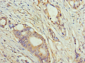
![TET1 antibody [N3C1] detects TET1 protein at nucleus in human A549 xenograft by immunohistochemical analysis. Sample: Paraffin-embedded human A549 xenograft . TET1 antibody [N3C1] (GTX124207) diluted at 1:250.
Antigen Retrieval: Trilogy? (EDTA based, pH 8.0) buffer, 15min](https://www.genetex.com/upload/website/prouct_img/normal/GTX124207/GTX124207_41479_20151227_IHC-P_w_23060522_200.webp "TET1 antibody [N3C1] detects TET1 protein at nucleus in human A549 xenograft by immunohistochemical analysis. Sample: Paraffin-embedded human A549 xenograft . TET1 antibody [N3C1] (GTX124207) diluted at 1:250.
Antigen Retrieval: Trilogy? (EDTA based, pH 8.0) buffer, 15min")
![TET1 antibody [N3C1] detects TET1 protein at nucleus by immunohistochemical analysis. Sample: Paraffin-embedded mouse testis. TET1 stained by TET1 antibody [N3C1] (GTX124207) diluted at 1:500. Antigen Retrieval: Citrate buffer, pH 6.0, 15 min](https://www.genetex.com/upload/website/prouct_img/normal/GTX124207/GTX124207_44531_20220311_IHC-P_M_w_23060522_765.webp "TET1 antibody [N3C1] detects TET1 protein at nucleus by immunohistochemical analysis. Sample: Paraffin-embedded mouse testis. TET1 stained by TET1 antibody [N3C1] (GTX124207) diluted at 1:500. Antigen Retrieval: Citrate buffer, pH 6.0, 15 min")
![TET1 antibody [N3C1] detects TET1 protein at nucleus by immunohistochemical analysis. Sample: Paraffin-embedded rat testis. TET1 stained by TET1 antibody [N3C1] (GTX124207) diluted at 1:500. Antigen Retrieval: Citrate buffer, pH 6.0, 15 min](https://www.genetex.com/upload/website/prouct_img/normal/GTX124207/GTX124207_44825_20230512_IHC-P_R_23060622_547.webp "TET1 antibody [N3C1] detects TET1 protein at nucleus by immunohistochemical analysis. Sample: Paraffin-embedded rat testis. TET1 stained by TET1 antibody [N3C1] (GTX124207) diluted at 1:500. Antigen Retrieval: Citrate buffer, pH 6.0, 15 min")
![TET1 antibody [N3C1] detects TET1 protein at nucleus by immunohistochemical analysis. Sample: Paraffin-embedded mouse testis. TET1 stained by TET1 antibody [N3C1] (GTX124207) diluted at 1:500. Antigen Retrieval: Citrate buffer, pH 6.0, 15 min](https://www.genetex.com/upload/website/prouct_img/normal/GTX124207/GTX124207_44825_20230512_IHC-P_M_23060622_910.webp "TET1 antibody [N3C1] detects TET1 protein at nucleus by immunohistochemical analysis. Sample: Paraffin-embedded mouse testis. TET1 stained by TET1 antibody [N3C1] (GTX124207) diluted at 1:500. Antigen Retrieval: Citrate buffer, pH 6.0, 15 min")
Non-transfected (–) and transfected (+) 293T whole cell extracts (50 μg) were separated by 5% SDS-PAGE, and the membrane was blotted with TET1 antibody [N3C1] (GTX124207) diluted at 1:1000. The HRP-conjugated anti-rabbit IgG antibody (GTX213110-01) was used to detect the primary antibody.
TET1 antibody [N3C1]
GTX124207
ApplicationsImmunoFluorescence, ImmunoPrecipitation, Western Blot, ChIP Chromatin ImmunoPrecipitation, ImmunoCytoChemistry, ImmunoHistoChemistry, ImmunoHistoChemistry Paraffin
Product group Antibodies
ReactivityHuman, Monkey, Mouse, Rat
TargetTET1
Overview
- SupplierGeneTex
- Product NameTET1 antibody [N3C1]
- Delivery Days Customer9
- Application Supplier NoteWB: 1:500-1:3000. ICC/IF: 1:100-1:1000. IHC-P: 1:100-1:1000. *Optimal dilutions/concentrations should be determined by the researcher.Not tested in other applications.
- ApplicationsImmunoFluorescence, ImmunoPrecipitation, Western Blot, ChIP Chromatin ImmunoPrecipitation, ImmunoCytoChemistry, ImmunoHistoChemistry, ImmunoHistoChemistry Paraffin
- CertificationResearch Use Only
- ClonalityPolyclonal
- Concentration1.01 mg/ml
- ConjugateUnconjugated
- Gene ID80312
- Target nameTET1
- Target descriptiontet methylcytosine dioxygenase 1
- Target synonymsCXXC6, LCX, bA119F7.1, methylcytosine dioxygenase TET1, CXXC finger 6, CXXC zinc finger 6, CXXC-type zinc finger protein 6, leukemia-associated protein with a CXXC domain, ten-eleven translocation 1 gene protein, ten-eleven translocation-1, tet oncogene 1
- HostRabbit
- IsotypeIgG
- Protein IDQ8NFU7
- Protein NameMethylcytosine dioxygenase TET1
- Scientific DescriptionDioxygenase that specifically binds methylcytosine (5mC), a minor base in mammalian DNA found in repetitive DNA elements that is crucial for retrotransposon silencing and mammalian development. Catalyzes the conversion of methylcytosine (5mC) to 5-hydroxymethylcytosine (hmC). The clear function of 5-hydroxymethylcytosine (hmC) is still unclear but it may influence chromatin structure and recruit specific factors or may constitute an intermediate component in cytosine demethylation. 5-hydroxymethylcytosine (hmC) is present in ES cells and is enriched in the brain, especially in Purkinje neurons. May play a role in the fetal development of heart, lung and brain.
- ReactivityHuman, Monkey, Mouse, Rat
- Storage Instruction-20°C or -80°C,2°C to 8°C
- UNSPSC41116161
Datasheet
Related products
Product group Antibodies
Anti-TET1 Antibody144-63563
ApplicationsImmunoFluorescence, Western Blot, ImmunoHistoChemistry
ReactivityHuman, Mouse, Rat
TargetTET1
- SizePrice
Product group Antibodies
Anti-TET1 AntibodyA13475
ApplicationsImmunoFluorescence, Western Blot, ImmunoCytoChemistry, ImmunoHistoChemistry
ReactivityHuman, Mouse, Rat
- SizePrice

Product group Antibodies
Anti-TET1 Antibody Picoband(r)A00603-2-CARRIER-FREE
ApplicationsFlow Cytometry, ImmunoFluorescence, Western Blot, ELISA, ImmunoCytoChemistry, ImmunoHistoChemistry
ReactivityHuman, Monkey
TargetTET1
- SizePrice
Product group Antibodies
TET1 AntibodyCSB-PA847709HA01HU
ApplicationsELISA, ImmunoHistoChemistry
ReactivityHuman
TargetTET1
- SizePrice
Product group Antibodies
Tet1 Polyclonal AntibodyCAC09616
ApplicationsELISA, ImmunoHistoChemistry
TargetTET1
- SizePrice
Product group Antibodies

LCX / TET1 AntibodyLS-C335161
ApplicationsImmunoPrecipitation, Western Blot
ReactivityHuman, Mouse, Rat
TargetTET1
- SizePrice
Product group Antibodies
References
TET1 antibodyGTX64332
ApplicationsImmunoFluorescence, Western Blot, ImmunoCytoChemistry, ImmunoHistoChemistry, ImmunoHistoChemistry Paraffin
ReactivityHuman, Mouse, Rat
TargetTET1
- SizePrice
![TET1 antibody [GT1462] detects TET1 protein at nucleus on HeLa xenograft by immunohistochemical analysis. Sample: Paraffin-embedded HeLa xenograft. TET1 antibody [GT1462] (GTX627420) dilution: 1:100.
Antigen Retrieval: Trilogy? (EDTA based, pH 8.0) buffer, 15min](https://www.genetex.com/upload/website/prouct_img/normal/GTX627420/GTX627420_40878_IHC_w_23061202_384.webp)
Product group Antibodies
TET1 antibody [GT1462]GTX627420
ApplicationsImmunoFluorescence, Western Blot, ImmunoCytoChemistry, ImmunoHistoChemistry, ImmunoHistoChemistry Paraffin
ReactivityHuman, Mouse
TargetTET1
- SizePrice
![TET1 antibody [GT465] detects TET1 protein by Western blot analysis. A. 30 μg 293T B. 30 μg of DDDDK-human TET1-transfected 293T cells 5 % SDS-PAGE TET1 antibody [GT465] (GTX629974) dilution: 1:1000](https://www.genetex.com/upload/website/prouct_img/normal/GTX629974/GTX629974_41491_WB_B_w_23061202_750.webp)
Product group Antibodies
TET1 antibody [GT465]GTX629974
ApplicationsImmunoFluorescence, Western Blot, ImmunoCytoChemistry
ReactivityHuman
TargetTET1
- SizePrice
![Various whole cell extracts (30 μg) were separated by 5% SDS-PAGE, and the membrane was blotted with TET1 antibody [GT1382] (GTX636643) diluted at 1:1000. The HRP-conjugated anti-mouse IgG antibody (GTX213111-01) was used to detect the primary antibody.](https://www.genetex.com/upload/website/prouct_img/normal/GTX636643/GTX636643_44566_20220121_WB_w_23051501_501.webp)
Product group Antibodies
TET1 antibody [GT1382]GTX636643
ApplicationsWestern Blot
ReactivityHuman
TargetTET1
- SizePrice