

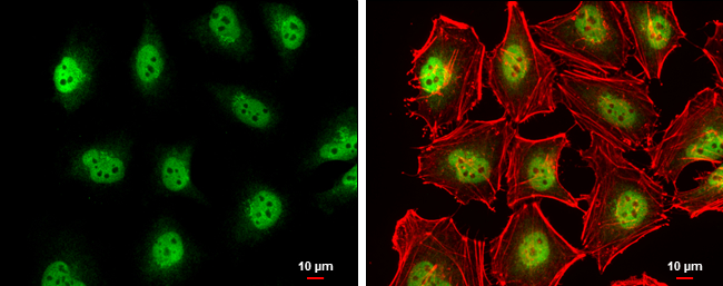
TET2 antibody [N2], N-term detects TET2 protein at nucleus by immunofluorescent analysis. Sample: HeLa cells were fixed in 4% paraformaldehyde at RT for 15 min. Green: TET2 protein stained by TET2 antibody [N2], N-term (GTX124204) diluted at 1:500. Red: phalloidin, a cytoskeleton marker, diluted at 1:200. Scale bar = 10 μm.
![Immunoprecipitation of TET2 protein from 293T whole cell extracts using 5 μg of TET2 antibody [N2], N-term (GTX124204). Western blot analysis was performed using TET2 antibody [N2], N-term (GTX124204) diluted at 1:500. EasyBlot HRP-conjugated anti rabbit IgG antibody (GTX221666-01) was used to detect the primary antibody.](https://www.genetex.com/upload/website/prouct_img/normal/GTX124204/GTX124204_41458_20170316_IP_w_23060522_930.webp "Immunoprecipitation of TET2 protein from 293T whole cell extracts using 5 μg of TET2 antibody [N2], N-term (GTX124204). Western blot analysis was performed using TET2 antibody [N2], N-term (GTX124204) diluted at 1:500. EasyBlot HRP-conjugated anti rabbit IgG antibody (GTX221666-01) was used to detect the primary antibody.")
 dilution: 1:5000 The HRP-conjugated anti-rabbit IgG antibody (GTX213110-01) was used to detect the primary antibody.")
![Wild-type (WT) and TET2 knockout (KO) HeLa cell extracts (30 μg) were separated by 5% SDS-PAGE, and the membrane was blotted with TET2 antibody [N2], N-term (GTX124204) diluted at 1:500. The HRP-conjugated anti-rabbit IgG antibody (GTX213110-01) was used to detect the primary antibody, and the signal was developed with Trident ECL plus-Enhanced.](https://www.genetex.com/upload/website/prouct_img/normal/GTX124204/GTX124204_41458_20181123_WB_KO_watermark_w_23060522_470.webp "Wild-type (WT) and TET2 knockout (KO) HeLa cell extracts (30 μg) were separated by 5% SDS-PAGE, and the membrane was blotted with TET2 antibody [N2], N-term (GTX124204) diluted at 1:500. The HRP-conjugated anti-rabbit IgG antibody (GTX213110-01) was used to detect the primary antibody, and the signal was developed with Trident ECL plus-Enhanced.")
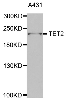
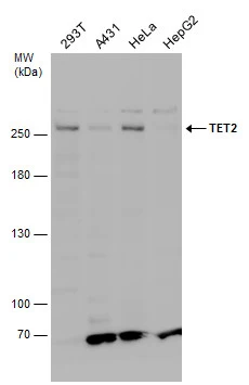
![Various whole cell extracts (30 μg) were separated by 5% SDS-PAGE, and the membrane was blotted with TET2 antibody [N2], N-term (GTX124204) diluted at 1:500. The HRP-conjugated anti-rabbit IgG antibody (GTX213110-01) was used to detect the primary antibody.](https://www.genetex.com/upload/website/prouct_img/normal/GTX124204/GTX124204_41458_20170309_WB_w_23060522_983.webp "Various whole cell extracts (30 μg) were separated by 5% SDS-PAGE, and the membrane was blotted with TET2 antibody [N2], N-term (GTX124204) diluted at 1:500. The HRP-conjugated anti-rabbit IgG antibody (GTX213110-01) was used to detect the primary antibody.")

TET2 antibody [N2], N-term detects TET2 protein at nucleus by immunofluorescent analysis. Sample: HeLa cells were fixed in 4% paraformaldehyde at RT for 15 min. Green: TET2 protein stained by TET2 antibody [N2], N-term (GTX124204) diluted at 1:500. Red: phalloidin, a cytoskeleton marker, diluted at 1:200. Scale bar = 10 μm.
TET2 antibody [N2], N-term
GTX124204
ApplicationsImmunoFluorescence, ImmunoPrecipitation, Western Blot, ChIP Chromatin ImmunoPrecipitation, ImmunoCytoChemistry
Product group Antibodies
ReactivityHuman
TargetTET2
Overview
- SupplierGeneTex
- Product NameTET2 antibody [N2], N-term
- Delivery Days Customer9
- Application Supplier NoteWB: 1:500-1:3000. ICC/IF: 1:100-1:1000. IP: 1:100-1:500. *Optimal dilutions/concentrations should be determined by the researcher.Not tested in other applications.
- ApplicationsImmunoFluorescence, ImmunoPrecipitation, Western Blot, ChIP Chromatin ImmunoPrecipitation, ImmunoCytoChemistry
- CertificationResearch Use Only
- ClonalityPolyclonal
- Concentration1 mg/ml
- ConjugateUnconjugated
- Gene ID54790
- Target nameTET2
- Target descriptiontet methylcytosine dioxygenase 2
- Target synonymsIMD75, KIAA1546, MDS, methylcytosine dioxygenase TET2, probable methylcytosine dioxygenase TET2, ten-eleven translocation 2, tet oncogene family member 2
- HostRabbit
- IsotypeIgG
- Protein IDQ6N021
- Protein NameMethylcytosine dioxygenase TET2
- ReactivityHuman
- Storage Instruction-20°C or -80°C,2°C to 8°C
- UNSPSC41116161
Datasheet
Related products
Product group Antibodies
Anti-TET2 AntibodyA30995
ApplicationsImmunoFluorescence, ImmunoPrecipitation, Western Blot
ReactivityHuman
- SizePrice
Product group Antibodies
Anti-TET2 Antibody144-63666
ApplicationsWestern Blot, ImmunoHistoChemistry
ReactivityHuman, Mouse, Rat
TargetTET2
- SizePrice

Product group Antibodies
Anti-TET2 AntibodyAMAB91439
ApplicationsWestern Blot, ImmunoCytoChemistry, ImmunoHistoChemistry
ReactivityHuman
TargetTET2
- SizePrice
Product group Antibodies
TET2 Polyclonal AntibodyBS-9449R
ApplicationsFlow Cytometry, ImmunoFluorescence, ImmunoCytoChemistry, ImmunoHistoChemistry, ImmunoHistoChemistry Frozen, ImmunoHistoChemistry Paraffin
ReactivityHuman, Mouse, Rat
TargetTET2
- SizePrice

Product group Antibodies
References

Goat anti-TET2EB09642
ApplicationsFlow Cytometry, ELISA, ImmunoHistoChemistry
ReactivityCanine, Human, Mouse
TargetTET2
- SizePrice
Product group Antibodies
TET2 AntibodyCSB-PA764560ESR1HU
ApplicationsELISA, ImmunoHistoChemistry
ReactivityHuman
TargetTET2
- SizePrice
Product group Antibodies
TET2 antibody, InternalGTX88329
ApplicationsImmunoHistoChemistry, ImmunoHistoChemistry Paraffin
ReactivityHuman
TargetTET2
- SizePrice
![TET2 antibody [N2-2], N-term detects TET2 protein at nucleus by immunohistochemical analysis. Sample: Paraffin-embedded human breast carcinoma. TET2 stained by TET2 antibody [N2-2], N-term (GTX124205) diluted at 1:500. Antigen Retrieval: Citrate buffer, pH 6.0, 15 min](https://www.genetex.com/upload/website/prouct_img/normal/GTX124205/GTX124205_44678_20220617_IHC-P_1_22062919_679.webp)
Product group Antibodies
TET2 antibody [N2-2], N-termGTX124205
ApplicationsImmunoFluorescence, ImmunoPrecipitation, Western Blot, ChIP Chromatin ImmunoPrecipitation, ImmunoCytoChemistry, ImmunoHistoChemistry, ImmunoHistoChemistry Paraffin
ReactivityHuman, Mouse
TargetTET2
- SizePrice
Product group Antibodies
TET2 antibodyGTX124227
ApplicationsWestern Blot
ReactivityHuman
TargetTET2
- SizePrice
![Wild-type (WT) and TET2 knockout (KO) HeLa cell extracts (30 μg) were separated by 5% SDS-PAGE, and the membrane was blotted with TET2 antibody [HL2822] (GTX640093) diluted at 1:1000. The HRP-conjugated anti-rabbit IgG antibody (GTX213110-01) was used to detect the primary antibody, and the signal was developed with Trident ECL plus-Enhanced.](https://www.genetex.com/upload/website/prouct_img/normal/GTX640093/GTX640093_T-45355_20240329_WB_KO_watermark_24040123_592.webp)
Product group Antibodies
TET2 antibody [HL2822]GTX640093
ApplicationsWestern Blot
ReactivityHuman
TargetTET2
- SizePrice