

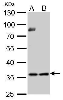
Thymidylate synthetase antibody detects TYMS protein by Western blot analysis. A. 30 μg PC-12 whole cell lysate/extract B. 30 μg Rat2 whole cell lysate/extract 10 % SDS-PAGE Thymidylate synthetase antibody (GTX103235) dilution: 1:1000
 A: Jurkat B: Raji C: THP-1 10% SDS PAGE GTX103235 diluted at 1:1000")
 dilution: 1:500.
Antigen Retrieval: Trilogy? (EDTA based, pH 8.0) buffer, 15min")
 dilution: 1:1000")
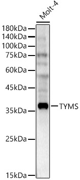
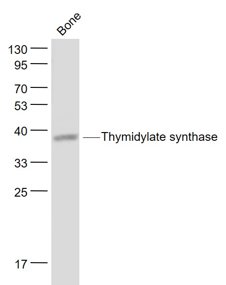
 were separated by 10% SDS-PAGE, and the membrane was blotted with Thymidylate synthetase antibody (GTX103235) diluted by 1:1000.")
![Thymidylate synthase antibody detects Thymidylate synthase protein at cytoplasm by immunofluorescent analysis. Sample: HeLa cells were fixed in 4% paraformaldehyde at RT for 15 min. Green: Thymidylate synthase stained by Thymidylate synthase antibody (GTX103235) diluted at 1:500. Red: alpha Tubulin, a cytoskeleton marker, stained by alpha Tubulin antibody [GT114] (GTX628802) diluted at 1:1000. Blue: Fluoroshield with DAPI (GTX30920).](https://www.genetex.com/upload/website/prouct_img/normal/GTX103235/GTX103235_44294_20220121_ICC_IF_w_23060119_748.webp "Thymidylate synthase antibody detects Thymidylate synthase protein at cytoplasm by immunofluorescent analysis. Sample: HeLa cells were fixed in 4% paraformaldehyde at RT for 15 min. Green: Thymidylate synthase stained by Thymidylate synthase antibody (GTX103235) diluted at 1:500. Red: alpha Tubulin, a cytoskeleton marker, stained by alpha Tubulin antibody [GT114] (GTX628802) diluted at 1:1000. Blue: Fluoroshield with DAPI (GTX30920).")
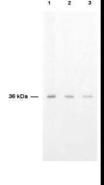
 10% SDS-PAGE The immunoprecipitated TYMS protein was detected by thymidylate synthetase antibody (GTX103235) diluted at 1 : 1000. EasyBlot anti-rabbit IgG (HRP) (GTX221666-01) was used as a secondary reagent.")
 dilution: 1:500.
Antigen Retrieval: Trilogy? (EDTA based, pH 8.0) buffer, 15min")
Thymidylate synthetase antibody detects TYMS protein by Western blot analysis. A. 30 μg PC-12 whole cell lysate/extract B. 30 μg Rat2 whole cell lysate/extract 10 % SDS-PAGE Thymidylate synthetase antibody (GTX103235) dilution: 1:1000
Thymidylate synthase antibody
GTX103235
ApplicationsImmunoFluorescence, ImmunoPrecipitation, Western Blot, ImmunoCytoChemistry, ImmunoHistoChemistry, ImmunoHistoChemistry Paraffin
Product group Antibodies
ReactivityHuman, Mouse, Rat
TargetTYMS
Overview
- SupplierGeneTex
- Product NameThymidylate synthase antibody
- Delivery Days Customer9
- Application Supplier NoteWB: 1:500-1:3000. ICC/IF: 1:100-1:1000. IHC-P: 1:100-1:1000. IP: 1:100-1:500. *Optimal dilutions/concentrations should be determined by the researcher.Not tested in other applications.
- ApplicationsImmunoFluorescence, ImmunoPrecipitation, Western Blot, ImmunoCytoChemistry, ImmunoHistoChemistry, ImmunoHistoChemistry Paraffin
- CertificationResearch Use Only
- ClonalityPolyclonal
- Concentration1 mg/ml
- ConjugateUnconjugated
- Gene ID7298
- Target nameTYMS
- Target descriptionthymidylate synthetase
- Target synonymsDKCD, HST422, TMS, TS, thymidylate synthase, TSase
- HostRabbit
- IsotypeIgG
- Protein IDP04818
- Protein NameThymidylate synthase
- Scientific DescriptionThymidylate synthase catalyzes the methylation of deoxyuridylate to deoxythymidylate using 5,10-methylenetetrahydrofolate (methylene-THF) as a cofactor. This function maintains the dTMP (thymidine-5-prime monophosphate) pool critical for DNA replication and repair. The enzyme has been of interest as a target for cancer chemotherapeutic agents. It is considered to be the primary site of action for 5-fluorouracil, 5-fluoro-2-prime-deoxyuridine, and some folate analogs. Expression of this gene and that of a naturally occuring antisense transcript rTSalpha (GeneID:55556) vary inversely when cell-growth progresses from late-log to plateau phase. [provided by RefSeq]
- ReactivityHuman, Mouse, Rat
- Storage Instruction-20°C or -80°C,2°C to 8°C
- UNSPSC41116161
Datasheet
Related products
Product group Antibodies
ApplicationsImmunoFluorescence, Western Blot, ImmunoCytoChemistry, ImmunoHistoChemistry
ReactivityHuman, Mouse, Rat
- SizePrice

Product group Antibodies
Anti-Thymidylate Synthase/TYMS Antibody Picoband(r)A04320-4-CARRIER-FREE
ApplicationsImmunoFluorescence, Western Blot, ELISA, ImmunoHistoChemistry
ReactivityHuman, Monkey, Rat
TargetTYMS
- SizePrice
Product group Antibodies
Anti-TYMS Antibody144-10441
ApplicationsImmunoFluorescence, Western Blot
ReactivityHuman, Mouse, Rat
TargetTYMS
- SizePrice
Product group Antibodies
TYMS Polyclonal AntibodyBS-8537R
ApplicationsImmunoFluorescence, Western Blot, ELISA, ImmunoCytoChemistry, ImmunoHistoChemistry, ImmunoHistoChemistry Frozen, ImmunoHistoChemistry Paraffin
ReactivityBovine, Human, Mouse, Porcine, Rabbit, Rat, Sheep
TargetTYMS
- SizePrice
Product group Antibodies
TYMS Polyclonal AntibodyCAC14768
ApplicationsImmunoFluorescence, ImmunoPrecipitation, Western Blot, ELISA
TargetTYMS
- SizePrice
Product group Antibodies
TYMS AntibodyCSB-PA025393LA01HU
ApplicationsImmunoFluorescence, ImmunoPrecipitation, Western Blot, ELISA
ReactivityHuman
TargetTYMS
- SizePrice
Product group Antibodies
Thymidylate synthase antibodyGTX27398
ApplicationsWestern Blot, ELISA
ReactivityHuman
TargetTYMS
- SizePrice
Product group Antibodies

TS / Thymidylate Synthase AntibodyLS-C497149
ApplicationsWestern Blot
ReactivityHuman, Mouse
TargetTYMS
- SizePrice

Product group Antibodies
Anti-TYMS AntibodyHPA074922
ApplicationsWestern Blot, ImmunoHistoChemistry
ReactivityHuman
TargetTYMS
- SizePrice
![Zebrafish tissue extract (30 μg) was separated by 12% SDS-PAGE, and the membrane was blotted with Thymidylate synthetase antibody [N3C3] (GTX113289) diluted at 1:500.](https://www.genetex.com/upload/website/prouct_img/normal/GTX113289/GTX113289_42480_20160519_WB_Z_eye_22111423_672.webp)
Product group Antibodies
Thymidylate synthase antibody [N3C3]GTX113289
ApplicationsWestern Blot
ReactivityHuman, Zebra Fish
TargetTYMS
- SizePrice