

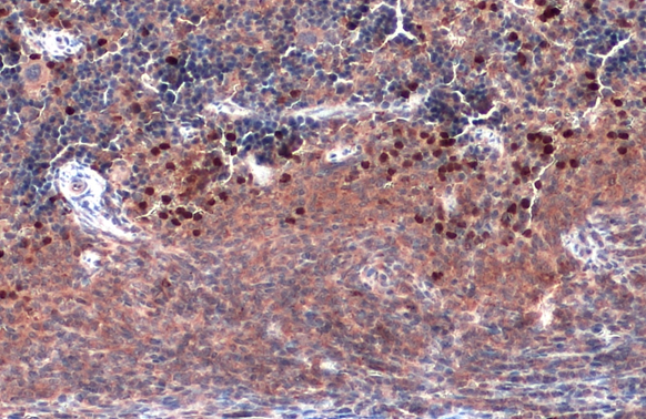
TLR9 antibody [N1N2], N-term detects TLR9 protein by immunohistochemical analysis. Sample: Paraffin-embedded mouse spleen. TLR9 stained by TLR9 antibody [N1N2], N-term (GTX111547) diluted at 1:500. Antigen Retrieval: Citrate buffer, pH 6.0, 15 min
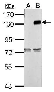
 A: Non-transfected 293T lysates B: TLR9 transfected 293T lysates 7.5% SDS PAGE GTX111547 diluted at 1:5000 The HRP-conjugated anti-rabbit IgG antibody (GTX213110-01) was used to detect the primary antibody.")
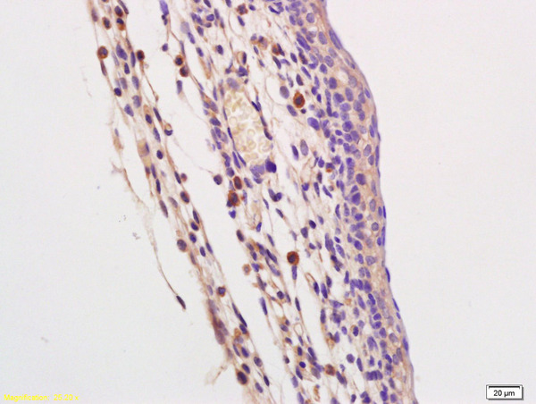
![TLR9 antibody [N1N2], N-term detects TLR9 protein by immunohistochemical analysis. Sample: Paraffin-embedded mouse placenta. TLR9 stained by TLR9 antibody [N1N2], N-term (GTX111547) diluted at 1:500. Antigen Retrieval: Citrate buffer, pH 6.0, 15 min](https://www.genetex.com/upload/website/prouct_img/normal/GTX111547/GTX111547_40079_20190412_IHC-P_M_1_w_23060500_342.webp "TLR9 antibody [N1N2], N-term detects TLR9 protein by immunohistochemical analysis. Sample: Paraffin-embedded mouse placenta. TLR9 stained by TLR9 antibody [N1N2], N-term (GTX111547) diluted at 1:500. Antigen Retrieval: Citrate buffer, pH 6.0, 15 min")
![TLR9 antibody [N1N2], N-term detects TLR9 protein by immunofluorescent analysis. Sample: HeLa cells were fixed in 4% paraformaldehyde for 10 min. Green: TLR9 protein stained by TLR9 antibody [N1N2], N-term (GTX111547) diluted at 1:100. Blue: Hoechst 33342 staining. Scale bar = 10 μm.](https://www.genetex.com/upload/website/prouct_img/normal/GTX111547/GTX111547_40079_IFA_w_23060500_779.webp "TLR9 antibody [N1N2], N-term detects TLR9 protein by immunofluorescent analysis. Sample: HeLa cells were fixed in 4% paraformaldehyde for 10 min. Green: TLR9 protein stained by TLR9 antibody [N1N2], N-term (GTX111547) diluted at 1:100. Blue: Hoechst 33342 staining. Scale bar = 10 μm.")
![Various whole cell extracts (30 μg) were separated by 5% SDS-PAGE, and the membrane was blotted with TLR9 antibody [N1N2], N-term (GTX111547) diluted at 1:1000. The HRP-conjugated anti-rabbit IgG antibody (GTX213110-01) was used to detect the primary antibody. Corresponding RNA expression data for the same cell lines are based on Human Protein Atlas program.](https://www.genetex.com/upload/website/prouct_img/normal/GTX111547/GTX111547_40079_20240112_WB_TPM_watermark_24011618_812.webp "Various whole cell extracts (30 μg) were separated by 5% SDS-PAGE, and the membrane was blotted with TLR9 antibody [N1N2], N-term (GTX111547) diluted at 1:1000. The HRP-conjugated anti-rabbit IgG antibody (GTX213110-01) was used to detect the primary antibody. Corresponding RNA expression data for the same cell lines are based on Human Protein Atlas program.")
![TLR9 antibody [N1N2], N-term detects TLR9 protein by immunofluorescent analysis. Sample: Mock and transfected 293T cells were fixed in 4% paraformaldehyde at RT for 15 min. Green: TLR9 stained by TLR9 antibody [N1N2], N-term (GTX111547) diluted at 1:500. Blue: Fluoroshield with DAPI (GTX30920).](https://www.genetex.com/upload/website/prouct_img/normal/GTX111547/GTX111547_40079_20240202_ICC_IF_B_24021917_830.webp "TLR9 antibody [N1N2], N-term detects TLR9 protein by immunofluorescent analysis. Sample: Mock and transfected 293T cells were fixed in 4% paraformaldehyde at RT for 15 min. Green: TLR9 stained by TLR9 antibody [N1N2], N-term (GTX111547) diluted at 1:500. Blue: Fluoroshield with DAPI (GTX30920).")
![Various tissue extracts (50 μg) were separated by 5% SDS-PAGE, and the membrane was blotted with TLR9 antibody [N1N2], N-term (GTX111547) diluted at 1:1000. The HRP-conjugated anti-rabbit IgG antibody (GTX213110-01) was used to detect the primary antibody, and the signal was developed with Trident ECL plus-Enhanced.](https://www.genetex.com/upload/website/prouct_img/normal/GTX111547/GTX111547_40079_20240308_WB_M_tissue_24031220_323.webp "Various tissue extracts (50 μg) were separated by 5% SDS-PAGE, and the membrane was blotted with TLR9 antibody [N1N2], N-term (GTX111547) diluted at 1:1000. The HRP-conjugated anti-rabbit IgG antibody (GTX213110-01) was used to detect the primary antibody, and the signal was developed with Trident ECL plus-Enhanced.")
TLR9 antibody [N1N2], N-term detects TLR9 protein by immunohistochemical analysis. Sample: Paraffin-embedded mouse spleen. TLR9 stained by TLR9 antibody [N1N2], N-term (GTX111547) diluted at 1:500. Antigen Retrieval: Citrate buffer, pH 6.0, 15 min
TLR9 antibody [N1N2], N-term
GTX111547
ApplicationsImmunoFluorescence, Western Blot, ImmunoCytoChemistry, ImmunoHistoChemistry, ImmunoHistoChemistry Paraffin
Product group Antibodies
ReactivityHuman, Mammals, Mouse
TargetTLR9
Overview
- SupplierGeneTex
- Product NameTLR9 antibody [N1N2], N-term
- Delivery Days Customer9
- Application Supplier NoteWB: 1:1000-1:10000. ICC/IF: 1:100-1:1000. IHC-P: 1:100-1:1000. *Optimal dilutions/concentrations should be determined by the researcher.Not tested in other applications.
- ApplicationsImmunoFluorescence, Western Blot, ImmunoCytoChemistry, ImmunoHistoChemistry, ImmunoHistoChemistry Paraffin
- CertificationResearch Use Only
- ClonalityPolyclonal
- Concentration0.71 mg/ml
- ConjugateUnconjugated
- Gene ID54106
- Target nameTLR9
- Target descriptiontoll like receptor 9
- Target synonymsCD289, toll-like receptor 9
- HostRabbit
- IsotypeIgG
- Protein IDQ9NR96
- Protein NameToll-like receptor 9
- Scientific DescriptionThe protein encoded by this gene is a member of the Toll-like receptor (TLR) family which plays a fundamental role in pathogen recognition and activation of innate immunity. TLRs are highly conserved from Drosophila to humans and share structural and functional similarities. They recognize pathogen-associated molecular patterns (PAMPs) that are expressed on infectious agents, and mediate the production of cytokines necessary for the development of effective immunity. The various TLRs exhibit different patterns of expression. This gene is preferentially expressed in immune cell rich tissues, such as spleen, lymph node, bone marrow and peripheral blood leukocytes. Studies in mice and human indicate that this receptor mediates cellular response to unmethylated CpG dinucleotides in bacterial DNA to mount an innate immune response. [provided by RefSeq]
- ReactivityHuman, Mammals, Mouse
- Storage Instruction-20°C or -80°C,2°C to 8°C
- UNSPSC12352203
References
- Kogan E, Berezovskiy Y, Blagova O, et al. Morphologically, immunohistochemically and PCR proven lymphocytic viral peri-, endo-, myocarditis in patients with fatal COVID-19. Diagn Pathol. 2022,17(1):31. doi: 10.1186/s13000-022-01207-6Read this paper
- Yanagawa M, Uchida K, Ando Y, et al. Basophils activated via TLR signaling may contribute to pathophysiology of type 1 autoimmune pancreatitis. J Gastroenterol. 2018,53(3):449-460. doi: 10.1007/s00535-017-1390-6Read this paper
- O'Hara JR, Feener TD, Fischer CD, et al. Campylobacter jejuni disrupts protective Toll-like receptor 9 signaling in colonic epithelial cells and increases the severity of dextran sulfate sodium-induced colitis in mice. Infect Immun. 2012,80(4):1563-71. doi: 10.1128/IAI.06066-11Read this paper
Datasheet
Related products

Product group Antibodies
Anti-TLR9 Antibody Picoband(r)A00198-3-CARRIER-FREE
ApplicationsWestern Blot, ImmunoHistoChemistry
ReactivityHuman, Rat
TargetTLR9
- SizePrice
Product group Antibodies
Anti-TLR9 Antibody144-63389
ApplicationsImmunoFluorescence, Western Blot
ReactivityHuman, Mouse, Rat
TargetTLR9
- SizePrice
Product group Antibodies
TLR9 Polyclonal AntibodyCAC13221
ApplicationsImmunoFluorescence, ELISA, ImmunoHistoChemistry
TargetTLR9
- SizePrice
Product group Antibodies
References
TLR9 Polyclonal AntibodyBS-2717R
ApplicationsFlow Cytometry, ImmunoFluorescence, Western Blot, ELISA, ImmunoCytoChemistry, ImmunoHistoChemistry, ImmunoHistoChemistry Frozen, ImmunoHistoChemistry Paraffin
ReactivityBovine, Canine, Equine, Human, Mouse, Porcine, Rat, Sheep
TargetTLR9
- SizePrice
Product group Antibodies
TLR9 AntibodyCSB-PA023608LA01HU
ApplicationsImmunoFluorescence, ELISA, ImmunoHistoChemistry
ReactivityHuman
TargetTLR9
- SizePrice
Product group Antibodies
TLR9 antibodyGTX31244
ApplicationsWestern Blot, ImmunoHistoChemistry, ImmunoHistoChemistry Paraffin
ReactivityHuman, Mouse
TargetTLR9
- SizePrice
Product group Antibodies
References
TLR9 antibody [5G5]GTX76032
ApplicationsFlow Cytometry, Western Blot, ImmunoHistoChemistry, ImmunoHistoChemistry Frozen, ImmunoHistoChemistry Paraffin
ReactivityCanine, Human, Mouse
TargetTLR9
- SizePrice
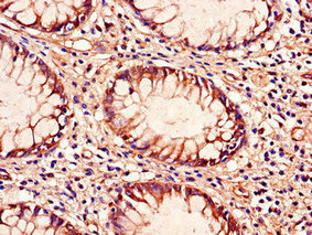
![TLR9 antibody [N2], N-term detects TLR9 protein by immunohistochemical analysis. Sample: Paraffin-embedded mouse spleen. TLR9 stained by TLR9 antibody [N2], N-term (GTX100726) diluted at 1:500. Antigen Retrieval: Citrate buffer, pH 6.0, 15 min](https://www.genetex.com/upload/website/prouct_img/normal/GTX100726/GTX100726_40016_20190412_IHC-P_M_w_23060100_421.webp)
Product group Antibodies
References
TLR9 antibody [N2], N-termGTX100726
ApplicationsWestern Blot, ImmunoHistoChemistry, ImmunoHistoChemistry Paraffin
ReactivityHuman, Mouse
TargetTLR9
- SizePrice
Product group Antibodies
TLR9 antibody [N3C2], InternalGTX111490
ApplicationsWestern Blot
ReactivityHuman
TargetTLR9
- SizePrice