

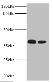
Western blot All lanes: Three-prime repair exonuclease 1 antibody at 3microg/ml Lane 1: Hela whole cell lysate Lane 2: HepG2 whole cell lysate Secondary Goat polyclonal to rabbit IgG at 1/10000 dilution Predicted band size: 39, 33, 34 kDa Observed band size: 39 kDa

 instead of CSB-PA865133ESR1HU in Hela whole cell lysate. For western blotting, a HRP-conjugated anti-rabbit IgG, specific to the non-reduced form of IgG was used as the Secondary antibody (1/50000) Lane 2: CSB-PA865133ESR1HU (4microg) + Hela whole cell lysate (500microg) Lane 3: Hela whole cell lysate (20microg)")
Western blot All lanes: Three-prime repair exonuclease 1 antibody at 3microg/ml Lane 1: Hela whole cell lysate Lane 2: HepG2 whole cell lysate Secondary Goat polyclonal to rabbit IgG at 1/10000 dilution Predicted band size: 39, 33, 34 kDa Observed band size: 39 kDa
TREX1 Antibody
CSB-PA865133ESR1HU
ApplicationsImmunoPrecipitation, Western Blot, ELISA, ImmunoHistoChemistry
Product group Antibodies
ReactivityHuman
TargetTREX1
Overview
- SupplierCusabio
- Product NameTREX1 Antibody
- Delivery Days Customer20
- ApplicationsImmunoPrecipitation, Western Blot, ELISA, ImmunoHistoChemistry
- CertificationResearch Use Only
- ClonalityPolyclonal
- ConjugateUnconjugated
- Gene ID11277
- Target nameTREX1
- Target descriptionthree prime repair exonuclease 1
- Target synonymsAGS1, CRV, DRN3, HERNS, RVCLS, three-prime repair exonuclease 1, 3' repair exonuclease 1, 3'-5' exonuclease TREX1, DNase III, deoxyribonuclease III
- HostRabbit
- IsotypeIgG
- Protein IDQ9NSU2
- Protein NameThree-prime repair exonuclease 1
- Scientific DescriptionMajor cellular 3-to-5 DNA exonuclease which digests single-stranded DNA (ssDNA) and double-stranded DNA (dsDNA) with mismatched 3 termini. Prevents cell-intrinsic initiation of autoimmunity. Acts by metabolizing DNA fragments from endogenous retroelements, including L1, LTR and SINE elements. Unless degraded, these DNA fragments accumulate in the cytosol and activate the IFN-stimulatory DNA (ISD) response and innate immune signaling. Prevents chronic ATM-dependent checkpoint activation, by processing ssDNA polynucleotide species arising from the processing of aberrant DNA replication intermediates. Inefficiently degrades oxidized DNA, such as that generated upon antimicrobial reactive oxygen production or upon absorption of UV light. During GZMA-mediated cell death, contributes to DNA damage in concert with NME1. NME1 nicks one strand of DNA and TREX1 removes bases from the free 3 end to enhance DNA damage and prevent DNA end reannealing and rapid repair.
- ReactivityHuman
- Storage Instruction-20°C or -80°C
- UNSPSC41116161
Related products
Product group Antibodies
Anti-TREX1 AntibodyA9994
ApplicationsImmunoFluorescence, Western Blot, ImmunoCytoChemistry
ReactivityHuman, Mouse
- SizePrice

Product group Antibodies
Anti-TREX1 AntibodyHPA035437
ApplicationsImmunoHistoChemistry
ReactivityHuman
TargetTREX1
- SizePrice
Product group Antibodies

TREX1 AntibodyLS-C335702
ApplicationsImmunoFluorescence, Western Blot
ReactivityHuman, Mouse
TargetTREX1
- SizePrice
Product group Antibodies
TREX1 Recombinant AntibodyBSM-61966R
ApplicationsImmunoFluorescence, Western Blot, ImmunoCytoChemistry, ImmunoHistoChemistry, ImmunoHistoChemistry Frozen, ImmunoHistoChemistry Paraffin
ReactivityHuman
TargetTREX1
- SizePrice
Product group Antibodies
Trex1 Polyclonal AntibodyCAC10572
ApplicationsImmunoPrecipitation, Western Blot, ELISA, ImmunoHistoChemistry
TargetTREX1
- SizePrice

Product group Antibodies
Anti-TREX1 Antibody Picoband(r)PB9748-CARRIER-FREE
ApplicationsWestern Blot
ReactivityHuman
TargetTREX1
- SizePrice
Product group Antibodies
TREX1 antibodyGTX114123
ApplicationsWestern Blot
ReactivityHuman
TargetTREX1
- SizePrice
Product group Antibodies
Anti-TREX1 Antibody144-06778
ApplicationsImmunoFluorescence, Western Blot
ReactivityHuman, Mouse
TargetTREX1
- SizePrice