

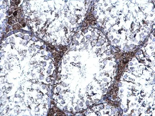
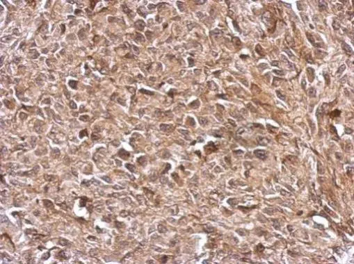
TrxR1 antibody [N1N3] detects TrxR1 protein at cytoplasm on mouse heart by immunohistochemical analysis. Sample: Paraffin-embedded mouse heart. TrxR1 antibody [N1N3] (GTX103202) diluted at 1:500.
Antigen Retrieval: Trilogy? (EDTA based, pH 8.0) buffer, 15min
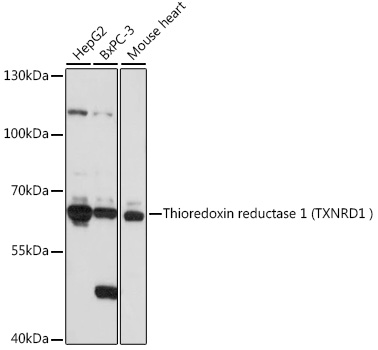
 A:Hep G2(GTX27900) 7.5% SDS PAGE GTX103202 diluted at 1:1000")
![TrxR1 antibody [N1N3] detects TrxR1 protein at cytoplasm on mouse heart by immunohistochemical analysis. Sample: Paraffin-embedded mouse heart. TrxR1 antibody [N1N3] (GTX103202) diluted at 1:500.
Antigen Retrieval: Trilogy? (EDTA based, pH 8.0) buffer, 15min](https://www.genetex.com/upload/website/prouct_img/normal/GTX103202/GTX103202_39862_20150116_IHC_M_2_w_23060119_225.webp "TrxR1 antibody [N1N3] detects TrxR1 protein at cytoplasm on mouse heart by immunohistochemical analysis. Sample: Paraffin-embedded mouse heart. TrxR1 antibody [N1N3] (GTX103202) diluted at 1:500.
Antigen Retrieval: Trilogy? (EDTA based, pH 8.0) buffer, 15min")
 antibody at 1:100 dilution.
Antigen Retrieval: Trilogy? (EDTA based, pH 8.0) buffer, 15min")
![TrxR1 antibody [N1N3] detects TrxR1 protein at nucleus by immunofluorescent analysis. Sample: MCF-7 cells were fixed in 4% paraformaldehyde at RT for 15 min. Green: TrxR1 protein stained by TrxR1 antibody [N1N3] (GTX103202) diluted at 1:500. Blue: Hoechst 33342 staining.](https://www.genetex.com/upload/website/prouct_img/normal/GTX103202/GTX103202_39862_IFA_w_23060119_904.webp "TrxR1 antibody [N1N3] detects TrxR1 protein at nucleus by immunofluorescent analysis. Sample: MCF-7 cells were fixed in 4% paraformaldehyde at RT for 15 min. Green: TrxR1 protein stained by TrxR1 antibody [N1N3] (GTX103202) diluted at 1:500. Blue: Hoechst 33342 staining.")
TrxR1 antibody [N1N3] detects TrxR1 protein at cytoplasm on mouse heart by immunohistochemical analysis. Sample: Paraffin-embedded mouse heart. TrxR1 antibody [N1N3] (GTX103202) diluted at 1:500.
Antigen Retrieval: Trilogy? (EDTA based, pH 8.0) buffer, 15min
TrxR1 antibody [N1N3]
GTX103202
ApplicationsImmunoFluorescence, Western Blot, ImmunoCytoChemistry, ImmunoHistoChemistry, ImmunoHistoChemistry Paraffin
Product group Antibodies
ReactivityHuman, Mouse
TargetTXNRD1
Overview
- SupplierGeneTex
- Product NameTrxR1 antibody [N1N3]
- Delivery Days Customer9
- Application Supplier NoteWB: 1:500-1:3000. ICC/IF: 1:100-1:1000. IHC-P: 1:100-1:1000. *Optimal dilutions/concentrations should be determined by the researcher.Not tested in other applications.
- ApplicationsImmunoFluorescence, Western Blot, ImmunoCytoChemistry, ImmunoHistoChemistry, ImmunoHistoChemistry Paraffin
- CertificationResearch Use Only
- ClonalityPolyclonal
- Concentration1 mg/ml
- ConjugateUnconjugated
- Gene ID7296
- Target nameTXNRD1
- Target descriptionthioredoxin reductase 1
- Target synonymsGRIM-12, TR, TR1, TRXR1, TXNR, TXNR1, thioredoxin reductase 1, cytoplasmic, KM-102-derived reductase-like factor, gene associated with retinoic and IFN-induced mortality 12 protein, gene associated with retinoic and interferon-induced mortality 12 protein, oxidoreductase, peroxidase TXNRD1, selenoprotein TXNRD1, testis tissue sperm-binding protein Li 46a, thioredoxin reductase GRIM-12, thioredoxin reductase TR1
- HostRabbit
- IsotypeIgG
- Protein IDQ16881
- Protein NameThioredoxin reductase 1, cytoplasmic
- Scientific DescriptionThis gene encodes a member of the family of pyridine nucleotide oxidoreductases. This protein reduces thioredoxins as well as other substrates, and plays a role in selenium metabolism and protection against oxidative stress. The functional enzyme is thought to be a homodimer which uses FAD as a cofactor. Each subunit contains a selenocysteine (Sec) residue which is required for catalytic activity. The selenocysteine is encoded by the UGA codon that normally signals translation termination. The 3 UTR of selenocysteine-containing genes have a common stem-loop structure, the sec insertion sequence (SECIS), that is necessary for the recognition of UGA as a Sec codon rather than as a stop signal. Alternative splicing results in several transcript variants encoding the same or different isoforms. [provided by RefSeq]
- ReactivityHuman, Mouse
- Storage Instruction-20°C or -80°C,2°C to 8°C
- UNSPSC41116161
Datasheet
Related products
Product group Antibodies
Anti-TXNRD1 AntibodyA90861
ApplicationsImmunoFluorescence, Western Blot, ImmunoCytoChemistry, ImmunoHistoChemistry
ReactivityHuman, Mouse, Rat
- SizePrice
Product group Antibodies
Anti-TXNRD1 Antibody144-64554
ApplicationsImmunoFluorescence, Western Blot, ImmunoHistoChemistry
ReactivityHuman, Mouse, Rat
TargetTXNRD1
- SizePrice

Product group Antibodies
Anti-TXNRD1 Antibody Picoband(r)A01778-3-CARRIER-FREE
ApplicationsWestern Blot, ELISA
ReactivityHuman
TargetTXNRD1
- SizePrice

Product group Antibodies
References
TXNRD1 Polyclonal AntibodyBS-8299R
ApplicationsFlow Cytometry, ImmunoFluorescence, ImmunoPrecipitation, Western Blot, ImmunoCytoChemistry, ImmunoHistoChemistry, ImmunoHistoChemistry Frozen, ImmunoHistoChemistry Paraffin
ReactivityBovine, Canine, Equine, Human, Mouse, Porcine, Rat
TargetTXNRD1
- SizePrice
Product group Antibodies

ApplicationsWestern Blot, ELISA, ImmunoHistoChemistry
ReactivityHuman
TargetTXNRD1
- SizePrice
Product group Antibodies
TXNRD1 AntibodyCSB-PA613705LA01HU
ApplicationsWestern Blot, ELISA, ImmunoHistoChemistry
ReactivityHuman
TargetTXNRD1
- SizePrice
Product group Antibodies
Txnrd1 Polyclonal AntibodyCAC09255
ApplicationsWestern Blot, ELISA, ImmunoHistoChemistry
TargetTXNRD1
- SizePrice
Product group Antibodies
TrxR1 antibody, C-termGTX13574
ApplicationsWestern Blot, ImmunoHistoChemistry, ImmunoHistoChemistry Paraffin
ReactivityHuman
TargetTXNRD1
- SizePrice
Product group Antibodies
TrxR1 antibodyGTX103207
ApplicationsImmunoFluorescence, Western Blot, ImmunoCytoChemistry, ImmunoHistoChemistry, ImmunoHistoChemistry Paraffin
ReactivityHuman, Mouse, Rat
TargetTXNRD1
- SizePrice
Product group Antibodies

TRXR1 / TXNRD1 Antibody (aa1-497)LS-C371361
ApplicationsELISA
ReactivityHuman
TargetTXNRD1
- SizePrice