


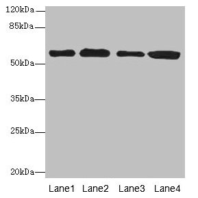
Figure 1. Western blot analysis of TXNRD1 using anti-TXNRD1 antibody (A01778-3). Electrophoresis was performed on a 5-20% SDS-PAGE gel at 70V (Stacking gel) / 90V (Resolving gel) for 2-3 hours. The sample well of each lane was loaded with 30 ug of sample under reducing conditions. Lane 1: human Jurkat whole cell lysates, Lane 2: human A549 whole cell lysates, Lane 3: human MCF-7 whole cell lysates, Lane 4: human Hela whole cell lysates. After electrophoresis, proteins were transferred to a nitrocellulose membrane at 150 mA for 50-90 minutes. Blocked the membrane with 5% non-fat milk/TBS for 1.5 hour at RT. The membrane was incubated with rabbit anti-TXNRD1 antigen affinity purified polyclonal antibody (Catalog # A01778-3) at 0.5 microg/mL overnight at 4°C, then washed with TBS-0.1%Tween 3 times with 5 minutes each and probed with a goat anti-rabbit IgG-HRP secondary antibody at a dilution of 1:5000 for 1.5 hour at RT. The signal is developed using an Enhanced Chemiluminescent detection (ECL) kit (Catalog # EK1002) with Tanon 5200 system. A specific band was detected for TXNRD1 at approximately 65 kDa. The expected band size for TXNRD1 is at 71 kDa.
Figure 1. Western blot analysis of TXNRD1 using anti-TXNRD1 antibody (A01778-3). Electrophoresis was performed on a 5-20% SDS-PAGE gel at 70V (Stacking gel) / 90V (Resolving gel) for 2-3 hours. The sample well of each lane was loaded with 30 ug of sample under reducing conditions. Lane 1: human Jurkat whole cell lysates, Lane 2: human A549 whole cell lysates, Lane 3: human MCF-7 whole cell lysates, Lane 4: human Hela whole cell lysates. After electrophoresis, proteins were transferred to a nitrocellulose membrane at 150 mA for 50-90 minutes. Blocked the membrane with 5% non-fat milk/TBS for 1.5 hour at RT. The membrane was incubated with rabbit anti-TXNRD1 antigen affinity purified polyclonal antibody (Catalog # A01778-3) at 0.5 microg/mL overnight at 4°C, then washed with TBS-0.1%Tween 3 times with 5 minutes each and probed with a goat anti-rabbit IgG-HRP secondary antibody at a dilution of 1:5000 for 1.5 hour at RT. The signal is developed using an Enhanced Chemiluminescent detection (ECL) kit (Catalog # EK1002) with Tanon 5200 system. A specific band was detected for TXNRD1 at approximately 65 kDa. The expected band size for TXNRD1 is at 71 kDa.
Anti-TXNRD1 Antibody Picoband(r)
A01778-3-CARRIER-FREE
ApplicationsWestern Blot, ELISA
Product group Antibodies
ReactivityHuman
TargetTXNRD1
Overview
- SupplierBoster Bio
- Product NameAnti-TXNRD1 Antibody Picoband(r)
- Delivery Days Customer9
- ApplicationsWestern Blot, ELISA
- CertificationResearch Use Only
- ClonalityPolyclonal
- Concentration500 ug/ml
- Gene ID7296
- Target nameTXNRD1
- Target descriptionthioredoxin reductase 1
- Target synonymsGRIM-12, TR, TR1, TRXR1, TXNR, TXNR1, thioredoxin reductase 1, cytoplasmic, KM-102-derived reductase-like factor, gene associated with retinoic and IFN-induced mortality 12 protein, gene associated with retinoic and interferon-induced mortality 12 protein, oxidoreductase, peroxidase TXNRD1, selenoprotein TXNRD1, testis tissue sperm-binding protein Li 46a, thioredoxin reductase GRIM-12, thioredoxin reductase TR1
- HostRabbit
- IsotypeIgG
- Protein IDQ16881
- Protein NameThioredoxin reductase 1, cytoplasmic
- Scientific DescriptionBoster Bio Anti-TXNRD1 Antibody Picoband® catalog # A01778-3. Tested in WB, ELISA applications. This antibody reacts with Human. The brand Picoband indicates this is a premium antibody that guarantees superior quality, high affinity, and strong signals with minimal background in Western blot applications. Only our best-performing antibodies are designated as Picoband, ensuring unmatched performance.
- ReactivityHuman
- Storage Instruction-20°C,2°C to 8°C
- UNSPSC12352203
Related products
Product group Antibodies
Anti-TXNRD1 AntibodyA90861
ApplicationsImmunoFluorescence, Western Blot, ImmunoCytoChemistry, ImmunoHistoChemistry
ReactivityHuman, Mouse, Rat
- SizePrice
Product group Antibodies
Anti-TXNRD1 Antibody144-64554
ApplicationsImmunoFluorescence, Western Blot, ImmunoHistoChemistry
ReactivityHuman, Mouse, Rat
TargetTXNRD1
- SizePrice

Product group Antibodies
References
TXNRD1 Polyclonal AntibodyBS-8299R
ApplicationsFlow Cytometry, ImmunoFluorescence, ImmunoPrecipitation, Western Blot, ImmunoCytoChemistry, ImmunoHistoChemistry, ImmunoHistoChemistry Frozen, ImmunoHistoChemistry Paraffin
ReactivityBovine, Canine, Equine, Human, Mouse, Porcine, Rat
TargetTXNRD1
- SizePrice
Product group Antibodies

ApplicationsWestern Blot, ELISA, ImmunoHistoChemistry
ReactivityHuman
TargetTXNRD1
- SizePrice
Product group Antibodies
TXNRD1 AntibodyCSB-PA613705LA01HU
ApplicationsWestern Blot, ELISA, ImmunoHistoChemistry
ReactivityHuman
TargetTXNRD1
- SizePrice
Product group Antibodies
Txnrd1 Polyclonal AntibodyCAC09255
ApplicationsWestern Blot, ELISA, ImmunoHistoChemistry
TargetTXNRD1
- SizePrice
![TrxR1 antibody [N1N3] detects TrxR1 protein at cytoplasm on mouse heart by immunohistochemical analysis. Sample: Paraffin-embedded mouse heart. TrxR1 antibody [N1N3] (GTX103202) diluted at 1:500.
Antigen Retrieval: Trilogy? (EDTA based, pH 8.0) buffer, 15min](https://www.genetex.com/upload/website/prouct_img/normal/GTX103202/GTX103202_39862_20150116_IHC_M_w_23060119_515.webp)
Product group Antibodies
TrxR1 antibody [N1N3]GTX103202
ApplicationsImmunoFluorescence, Western Blot, ImmunoCytoChemistry, ImmunoHistoChemistry, ImmunoHistoChemistry Paraffin
ReactivityHuman, Mouse
TargetTXNRD1
- SizePrice
Product group Antibodies

TRXR1 / TXNRD1 Antibody (aa1-497)LS-C371361
ApplicationsELISA
ReactivityHuman
TargetTXNRD1
- SizePrice

Product group Antibodies
Anti-TXNRD1 AntibodyHPA001395
ApplicationsWestern Blot, ImmunoHistoChemistry
ReactivityHuman
TargetTXNRD1
- SizePrice
Product group Antibodies
TargetTXNRD1
- SizePrice