


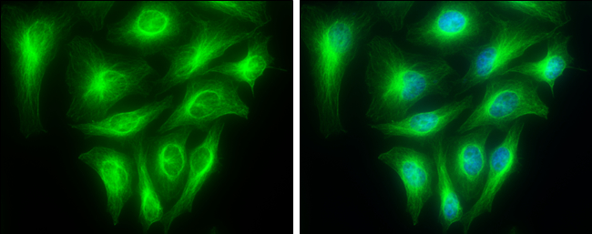
Confocal microscopy of PFA-fixed and 0.15% Triton X-100 permealised HeLa cells stained with TUBA1A Rabbit Recombinant Antibody AE00320 at 10ug/ml for 1h at RT. Detection by CF488 (green) for the antibody and DAPI (blue) for nuclear staining. Bottom right shows staining with an isotype control antibody.

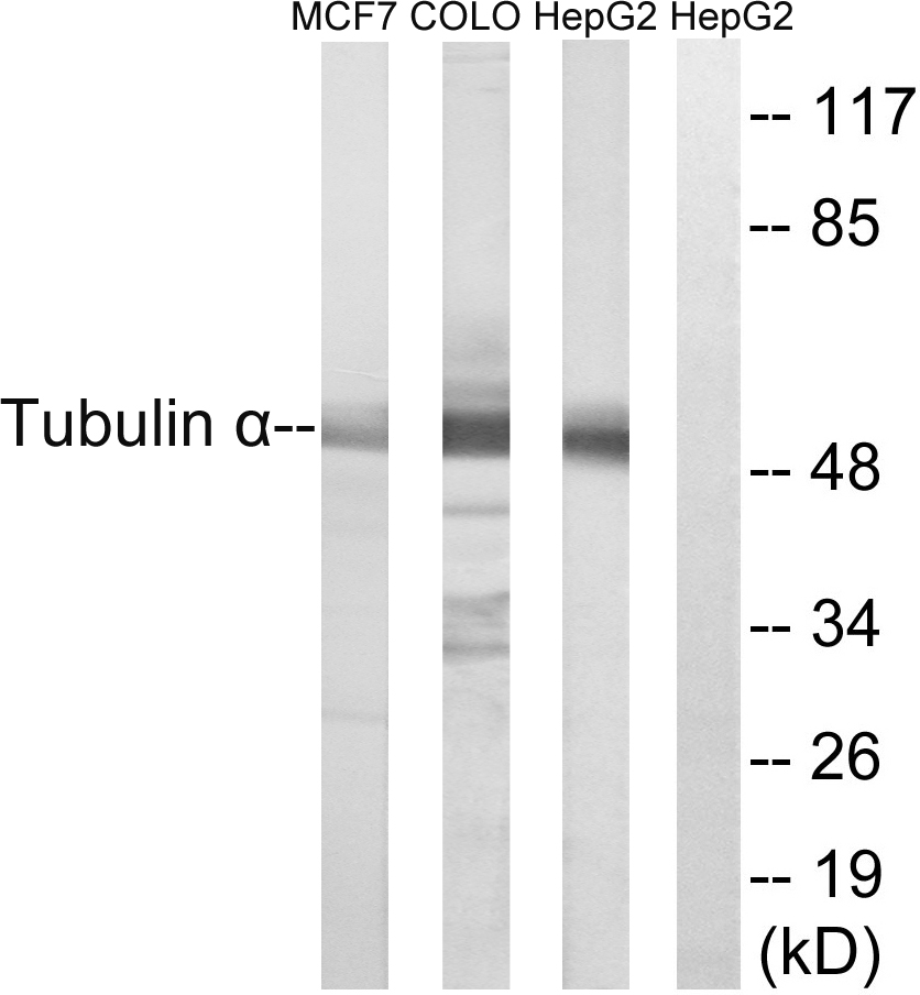
 from HeLa (A), HEK293 (B) and HepG2 (C) stained with TUBA1A Rabbit Recombinant Antibody AE00320 at 0.01ug/ml (1h at ambient temp). ECL staining by HRP.")
Confocal microscopy of PFA-fixed and 0.15% Triton X-100 permealised HeLa cells stained with TUBA1A Rabbit Recombinant Antibody AE00320 at 10ug/ml for 1h at RT. Detection by CF488 (green) for the antibody and DAPI (blue) for nuclear staining. Bottom right shows staining with an isotype control antibody.
TUBA1A Recombinant Antibody AE00320
AE00320
TargetTUBA1A
Product group Antibodies
Product AE00320 is not available
Product not available
There may be an alternative product available, please contact our technical support team.
Overview
- SupplierAeonian Biotech
- Product NameTUBA1A Recombinant Antibody AE00320
- Delivery Days Customer9
- Applications SupplierICC, IHC, WB
- CertificationResearch Use Only
- ClonalityRecombinant
- Clone IDF2CR
- ConjugateUnconjugated
- Gene ID7846
- Target nameTUBA1A
- Target descriptiontubulin alpha 1a
- Target synonymsB-ALPHA-1, LIS3, TUBA3, tubulin alpha-1A chain, hum-a-tub1, hum-a-tub2, tubulin B-alpha-1, tubulin alpha-3 chain, tubulin, alpha, brain-specific
- HostRabbit
- IsotypeIgG lambda
- Protein IDQ71U36
- Protein NameTubulin alpha-1A chain
- Scientific DescriptionTUBA1A Recombinant Antibody AE00320
- Shelf life instructionIntegrity warranted for 24 months after purchase when handled and stored according to instructions, see below.
- Reactivity SupplierHuman
- Storage Instruction2-8°C
- UNSPSC12352203
Datasheet
MSDS
Related products
Product group Antibodies
Anti-Tubulin alpha Antibody102-25843
ApplicationsImmunoFluorescence, Western Blot, ImmunoHistoChemistry
TargetTUBA1A
- SizePrice
Product group Antibodies

Anti-Alpha-Tubulin [F2C]Ab00403-1.1
ApplicationsImmunoFluorescence, Western Blot, ELISA
ReactivityHuman
TargetTUBA1A
- SizePrice

Product group Antibodies
Anti-Tubulin alpha Antibody Picoband(r)A03989-1-CARRIER-FREE
ApplicationsFlow Cytometry, ImmunoFluorescence, Western Blot, ELISA, ImmunoCytoChemistry, ImmunoHistoChemistry
ReactivityHuman, Mouse, Rat
TargetTUBA1A
- SizePrice
Product group Antibodies
References
alpha Tubulin 1A antibodyGTX109832
ApplicationsImmunoFluorescence, Western Blot, ImmunoCytoChemistry, ImmunoHistoChemistry, ImmunoHistoChemistry Paraffin
ReactivityDrosophila, Human, Mouse, Rat
TargetTUBA1A
- SizePrice
Product group Antibodies
TUBA1A Monoclonal AntibodyCAC12980
ApplicationsFlow Cytometry, ImmunoFluorescence, ImmunoPrecipitation, Western Blot, ELISA, ImmunoHistoChemistry
ReactivityMouse, Rabbit, Rat
- SizePrice
Product group Antibodies
Tubulin alpha Polyclonal AntibodyBS-20496R
ApplicationsImmunoFluorescence, Western Blot, ELISA, ImmunoCytoChemistry, ImmunoHistoChemistry, ImmunoHistoChemistry Frozen, ImmunoHistoChemistry Paraffin
ReactivityHuman, Mouse, Rat
TargetTUBA1A
- SizePrice
Product group Antibodies
ApplicationsWestern Blot
ReactivityHuman
TargetTUBA1A
- SizePrice
Product group Antibodies
ApplicationsImmunoFluorescence, Western Blot, ELISA, ImmunoHistoChemistry
ReactivityHuman, Mouse, Rat
- SizePrice

Product group Antibodies
Anti-TUBA1A AntibodyHPA039247
ApplicationsWestern Blot, ImmunoCytoChemistry, ImmunoHistoChemistry
ReactivityHuman, Mouse, Rat
TargetTUBA1A
- SizePrice
Product group Antibodies
ApplicationsWestern Blot, ELISA
ReactivityHuman, Mouse, Rat
TargetTUBA1A
- SizePrice