

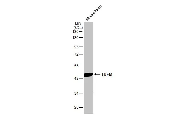
Mouse tissue extract (50 μg) was separated by 10% SDS-PAGE, and the membrane was blotted with TUFM antibody [HL2117] (GTX638089) diluted at 1:5000. The HRP-conjugated anti-rabbit IgG antibody (GTX213110-01) was used to detect the primary antibody.
![Rat tissue extract (50 μg) was separated by 10% SDS-PAGE, and the membrane was blotted with TUFM antibody [HL2117] (GTX638089) diluted at 1:5000. The HRP-conjugated anti-rabbit IgG antibody (GTX213110-01) was used to detect the primary antibody.](https://www.genetex.com/upload/website/prouct_img/normal/GTX638089/GTX638089_T-44914_20230203_WB_R_muscle_23020621_246.webp "Rat tissue extract (50 μg) was separated by 10% SDS-PAGE, and the membrane was blotted with TUFM antibody [HL2117] (GTX638089) diluted at 1:5000. The HRP-conjugated anti-rabbit IgG antibody (GTX213110-01) was used to detect the primary antibody.")
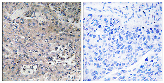
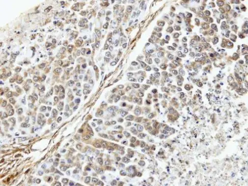
![TUFM antibody [HL2117] detects TUFM protein at mitochondria by immunohistochemical analysis. Sample: Paraffin-embedded mouse heart. TUFM stained by TUFM antibody [HL2117] (GTX638089) diluted at 1:100. Antigen Retrieval: Citrate buffer, pH 6.0, 15 min](https://www.genetex.com/upload/website/prouct_img/normal/GTX638089/GTX638089_T-44914_20230203_IHC-P_M_23021401_969.webp "TUFM antibody [HL2117] detects TUFM protein at mitochondria by immunohistochemical analysis. Sample: Paraffin-embedded mouse heart. TUFM stained by TUFM antibody [HL2117] (GTX638089) diluted at 1:100. Antigen Retrieval: Citrate buffer, pH 6.0, 15 min")
![TUFM antibody [HL2117] detects TUFM protein at mitochondria by immunohistochemical analysis. Sample: Paraffin-embedded rat kidney. TUFM stained by TUFM antibody [HL2117] (GTX638089) diluted at 1:100. Antigen Retrieval: Citrate buffer, pH 6.0, 15 min](https://www.genetex.com/upload/website/prouct_img/normal/GTX638089/GTX638089_T-44914_20230203_IHC-P_R_23021401_865.webp "TUFM antibody [HL2117] detects TUFM protein at mitochondria by immunohistochemical analysis. Sample: Paraffin-embedded rat kidney. TUFM stained by TUFM antibody [HL2117] (GTX638089) diluted at 1:100. Antigen Retrieval: Citrate buffer, pH 6.0, 15 min")
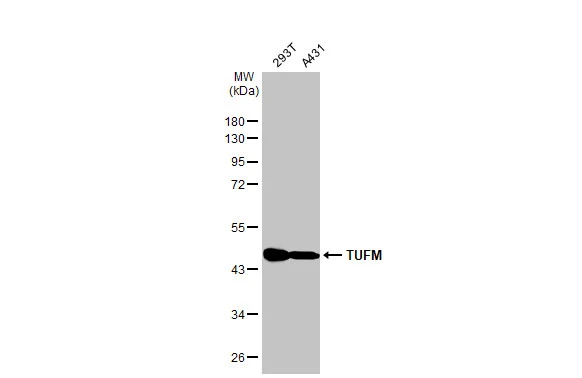
![Various whole cell extracts (30 μg) were separated by 10% SDS-PAGE, and the membrane was blotted with TUFM antibody [HL2117] (GTX638089) diluted at 1:5000. The HRP-conjugated anti-rabbit IgG antibody (GTX213110-01) was used to detect the primary antibody.](https://www.genetex.com/upload/website/prouct_img/normal/GTX638089/GTX638089_44984_20230317_WB_23032022_953.webp "Various whole cell extracts (30 μg) were separated by 10% SDS-PAGE, and the membrane was blotted with TUFM antibody [HL2117] (GTX638089) diluted at 1:5000. The HRP-conjugated anti-rabbit IgG antibody (GTX213110-01) was used to detect the primary antibody.")
![Whole zebrafish extract (30 μg) was separated by 10% SDS-PAGE, and the membrane was blotted with TUFM antibody [HL2117] (GTX638089) diluted at 1:1000. The HRP-conjugated anti-rabbit IgG antibody (GTX213110-01) was used to detect the primary antibody.](https://www.genetex.com/upload/website/prouct_img/normal/GTX638089/GTX638089_44984_20230609_WB_Z_23061400_318.webp "Whole zebrafish extract (30 μg) was separated by 10% SDS-PAGE, and the membrane was blotted with TUFM antibody [HL2117] (GTX638089) diluted at 1:1000. The HRP-conjugated anti-rabbit IgG antibody (GTX213110-01) was used to detect the primary antibody.")
Mouse tissue extract (50 μg) was separated by 10% SDS-PAGE, and the membrane was blotted with TUFM antibody [HL2117] (GTX638089) diluted at 1:5000. The HRP-conjugated anti-rabbit IgG antibody (GTX213110-01) was used to detect the primary antibody.
TUFM antibody [HL2117]
GTX638089
ApplicationsWestern Blot, ImmunoHistoChemistry, ImmunoHistoChemistry Paraffin
Product group Antibodies
ReactivityHuman, Mouse, Rat, Zebra Fish
TargetTUFM
Overview
- SupplierGeneTex
- Product NameTUFM antibody [HL2117]
- Delivery Days Customer9
- Application Supplier NoteWB: 1:1000-1:10000. *Optimal dilutions/concentrations should be determined by the researcher.Not tested in other applications.
- ApplicationsWestern Blot, ImmunoHistoChemistry, ImmunoHistoChemistry Paraffin
- CertificationResearch Use Only
- ClonalityMonoclonal
- Clone IDHL2117
- Concentration1 mg/ml
- ConjugateUnconjugated
- Gene ID7284
- Target nameTUFM
- Target descriptionTu translation elongation factor, mitochondrial
- Target synonymsCOXPD4, EF-TuMT, EFTU, P43, elongation factor Tu, mitochondrial, EF-Tu, epididymis secretory sperm binding protein
- HostRabbit
- IsotypeIgG
- Protein IDP49411
- Protein NameElongation factor Tu, mitochondrial
- Scientific DescriptionThis gene encodes a protein which participates in protein translation in mitochondria. Mutations in this gene have been associated with combined oxidative phosphorylation deficiency resulting in lactic acidosis and fatal encephalopathy. A pseudogene has been identified on chromosome 17. [provided by RefSeq, Jul 2008]
- ReactivityHuman, Mouse, Rat, Zebra Fish
- Storage Instruction-20°C or -80°C,2°C to 8°C
- UNSPSC41116161
Datasheet
Related products
Product group Antibodies
Anti-TUFM AntibodyA97759
ApplicationsELISA, ImmunoHistoChemistry
ReactivityHuman, Mouse, Rat
- SizePrice

Product group Antibodies
Anti-TUFM Antibody Picoband(r)A05606-2-CARRIER-FREE
ApplicationsImmunoFluorescence, Western Blot, ELISA, ImmunoCytoChemistry
ReactivityHuman, Mouse, Rat
TargetTUFM
- SizePrice
Product group Antibodies
Anti-TUFM Antibody144-06423
ApplicationsImmunoFluorescence, ImmunoPrecipitation, Western Blot, ImmunoHistoChemistry
ReactivityHuman, Mouse, Rat
TargetTUFM
- SizePrice

Product group Antibodies
Anti-TUFM AntibodyAMAB90964
ApplicationsWestern Blot, ImmunoCytoChemistry, ImmunoHistoChemistry
ReactivityHuman
TargetTUFM
- SizePrice
Product group Antibodies
TUFM Polyclonal AntibodyCAC13937
ApplicationsWestern Blot, ELISA
TargetTUFM
- SizePrice
Product group Antibodies
TUFM AntibodyCSB-PA02199A0RB
ApplicationsWestern Blot, ELISA
ReactivityHuman
TargetTUFM
- SizePrice
Product group Antibodies
TUFM antibodyGTX101763
ApplicationsImmunoFluorescence, ImmunoPrecipitation, Western Blot, ImmunoCytoChemistry, ImmunoHistoChemistry, ImmunoHistoChemistry Paraffin
ReactivityHuman, Mouse
TargetTUFM
- SizePrice
Product group Antibodies
TUFM antibody [N3C3]GTX101764
ApplicationsImmunoPrecipitation, Western Blot, ImmunoHistoChemistry, ImmunoHistoChemistry Paraffin, Other Application
ReactivityHuman
TargetTUFM
- SizePrice
![TUFM antibody [HL2193] detects TUFM protein at mitochondria by immunofluorescent analysis. Sample: HeLa cells were fixed in 4% paraformaldehyde at RT for 15 min. Green: TUFM stained by TUFM antibody [HL2193] (GTX638197) diluted at 1:500. Red: alpha Tubulin, a cytoskeleton marker, stained by alpha Tubulin antibody [GT114] (GTX628802) diluted at 1:1000. Blue: Fluoroshield with DAPI (GTX30920).](https://www.genetex.com/upload/website/prouct_img/normal/GTX638197/GTX638197_T-44942_20230331_ICC_IF_23041023_194.webp)
Product group Antibodies
TUFM antibody [HL2193]GTX638197
ApplicationsImmunoFluorescence, Western Blot, ImmunoCytoChemistry
ReactivityHuman
TargetTUFM
- SizePrice
Product group Antibodies
TUFM antibodyGTX55825
ApplicationsImmunoFluorescence, ImmunoPrecipitation, Western Blot, ImmunoCytoChemistry, ImmunoHistoChemistry, ImmunoHistoChemistry Paraffin
ReactivityHuman, Mouse, Rat
TargetTUFM
- SizePrice