

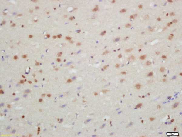
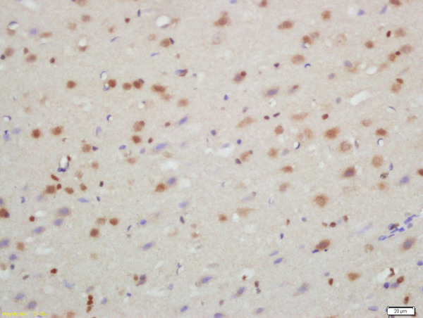
IHC-P analysis of rat brain tissue using GTX57221 TXNIP antibody. Dilution : 1:200
IHC-P analysis of rat brain tissue using GTX57221 TXNIP antibody. Dilution : 1:200
TXNIP antibody
GTX57221
ApplicationsImmunoFluorescence, Western Blot, ImmunoCytoChemistry, ImmunoHistoChemistry, ImmunoHistoChemistry Paraffin
Product group Antibodies
ReactivityHuman, Mouse, Rat
TargetTXNIP
Overview
- SupplierGeneTex
- Product NameTXNIP antibody
- Delivery Days Customer9
- Application Supplier NoteWB: 1:300-1:1000. IHC-P: 1:50-400. *Optimal dilutions/concentrations should be determined by the researcher.Not tested in other applications.
- ApplicationsImmunoFluorescence, Western Blot, ImmunoCytoChemistry, ImmunoHistoChemistry, ImmunoHistoChemistry Paraffin
- CertificationResearch Use Only
- ClonalityPolyclonal
- Concentration1 mg/ml
- ConjugateUnconjugated
- Gene ID10628
- Target nameTXNIP
- Target descriptionthioredoxin interacting protein
- Target synonymsARRDC6, EST01027, HHCPA78, THIF, VDUP1, thioredoxin-interacting protein, thioredoxin binding protein 2, upregulated by 1,25-dihydroxyvitamin D-3, vitamin D3 up-regulated protein 1
- HostRabbit
- IsotypeIgG
- Protein IDQ9H3M7
- Protein NameThioredoxin-interacting protein
- Scientific DescriptionThis gene encodes a thioredoxin-binding protein that is a member of the alpha arrestin protein family. Thioredoxin is a thiol-oxidoreductase that is a major regulator of cellular redox signaling which protects cells from oxidative stress. This protein inhibits the antioxidative function of thioredoxin resulting in the accumulation of reactive oxygen species and cellular stress. This protein also functions as a regulator of cellular metabolism and of endoplasmic reticulum (ER) stress. This protein may also function as a tumor suppressor. Alternate splicing results in multiple transcript variants. [provided by RefSeq, Sep 2015]
- ReactivityHuman, Mouse, Rat
- Storage Instruction-20°C or -80°C,2°C to 8°C
- UNSPSC12352203
Datasheet
Related products
Product group Antibodies
Txnip Polyclonal AntibodyCAC11357
ApplicationsImmunoFluorescence, ELISA
TargetTXNIP
- SizePrice
Product group Antibodies
References
TXNIP Polyclonal AntibodyBS-3897R
ApplicationsFlow Cytometry, ImmunoFluorescence, Western Blot, ELISA, ImmunoCytoChemistry, ImmunoHistoChemistry, ImmunoHistoChemistry Frozen, ImmunoHistoChemistry Paraffin
ReactivityCanine, Human, Mouse, Porcine, Rabbit, Rat
TargetTXNIP
- SizePrice
Product group Antibodies
Anti-TXNIP AntibodyA81009
ApplicationsImmunoFluorescence, Western Blot, ImmunoCytoChemistry
ReactivityHuman, Mouse, Rat
- SizePrice
Product group Antibodies
Anti-TXNIP Antibody144-66751
ApplicationsWestern Blot
ReactivityHuman, Mouse
TargetTXNIP
- SizePrice
Product group Antibodies
TXNIP antibodyGTX31592
ApplicationsWestern Blot, ELISA, ImmunoHistoChemistry, ImmunoHistoChemistry Paraffin
ReactivityHuman, Mouse, Rat
TargetTXNIP
- SizePrice
Product group Antibodies

TXNIP AntibodyLS-C771531
ApplicationsELISA, ImmunoHistoChemistry
ReactivityHuman, Mouse, Rat
TargetTXNIP
- SizePrice
Product group Antibodies
TXNIP antibody, C-termGTX45803
ApplicationsWestern Blot
ReactivityHuman
TargetTXNIP
- SizePrice