

VEGF Receptor 2 antibody [260.4(KDR-2)] (Biotin)
GTX10975
ApplicationsFlow Cytometry, Western Blot
Product group Antibodies
TargetKDR
Overview
- SupplierGeneTex
- Product NameVEGF Receptor 2 antibody [260.4(KDR-2)] (Biotin)
- Delivery Days Customer9
- Application Supplier NoteWB: 1:1,000. *Optimal dilutions/concentrations should be determined by the researcher.Not tested in other applications.
- ApplicationsFlow Cytometry, Western Blot
- CertificationResearch Use Only
- ClonalityMonoclonal
- Clone ID260.4(KDR-2)
- ConjugateBiotin
- Gene ID3791
- Target nameKDR
- Target descriptionkinase insert domain receptor
- Target synonymsCD309, FLK1, VEGFR, VEGFR2, vascular endothelial growth factor receptor 2, fetal liver kinase-1, kinase insert domain receptor (a type III receptor tyrosine kinase), protein-tyrosine kinase receptor Flk-1, soluble VEGFR2, tyrosine kinase growth factor receptor
- HostMouse
- IsotypeIgG1
- Protein IDP35968
- Protein NameVascular endothelial growth factor receptor 2
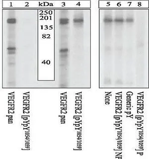
- Scientific DescriptionVascular endothelial growth factor (VEGF), also called vasculotropin (VAS) and vascular permeability factor (VPF), is a member of a family of endothelial cell mitogens and angiogenic factors. VEGF is a homodimeric heparin-binding glycoprotein, which specifically stimulates the proliferation of endothelial cells isolated from both small and large vessels. These include endothelial cells from adrenal cortex, cerebral cortex, fetal and adult aorta and human umbilical vein. The mitogenic activity of VEGF appears to be stimulated by specific VEGF receptors (160-200 kDa) which can be found on the surface of various endothelial cells. VEGF binds to two structurally similar receptor tyrosine kinases; Flt17(fms-like tyrosine kinase 1, also known as VEGF Receptor 1), and KDR8 (kinase-insert domain containing receptor, also known as VEGF Receptor 2). The homologous gene for the human KDR gene is mouse Flk1 (fetal liver kinase 1) and rat TKrc Both receptors are members of a superfamily of receptor tyrosine kinases (RTKs) which include PDGF receptor, c-fms, M-CSF receptor and c-kit - the receptor for stem-cell factor. The KDR gene has been mapped to human chromosome 4q11-q12, which is the same locus of PDGF receptor and c-kit. The extracellular domains of KDR/Flk1 and Flt1 (approx. 90 kDa), have seven immunoglobulin-like domains and belong to the class III RTKs. This family includes also Flt4, which shares a high homology with the two VEGF receptors. Studies using KDR and Flt1 stably transfected endothelial cell lines have shown that these two receptors exhibit different affinities to VEGF and mediate different responses. KDR expressing cells show striking changes in cell morphology, actin reorganization and membrane ruffling, chemotaxis and mitogenicity in response to VEGF, while Flt1 expressing cells lack such responses. Both KDR and Flt1 are phosphorylated in response to VEGF, however KDR much more efficiently. KDR/Flk1 does not respond to placental growth factor (PlGF), a VEGF related growth factor, while Flt1 binds PlGF specifically. The expression pattern of the two receptors is somewhat different; Flt1 is predominately expressed in human placenta and human vascular endothelial cells, while KDR is more widely expressed in all vessel-derived endothelial cells but low in human and fetal bovine placenta. Both VEGF receptors (KDR and Flt1) are upregulated in human fetal and in adult kidney. It has been suggested that in human postnatal hematopoietic tissues, 0.1% to 0.5% of CD34+ cells express VEGF Receptor 2/KDR; pluripotent hematopoietic stem cells (HSCs) were found to be restricted to the CD34+KDR+ cell fraction, while lineage-committed hematopoietic progenitor cells (HPCs) were found in the CD34+KDR- subset. Antibodies that react specifically with VEGF receptors are useful for the study of the specific differential tissue expression and intracellular localization of the receptor in normal and neoplastic tissue.
- Storage Instruction-20°C or -80°C,2°C to 8°C
- UNSPSC12352203
Datasheet
Related products
Product group Antibodies
KDR AntibodyCSB-PA002542
ApplicationsImmunoFluorescence, Western Blot, ELISA, ImmunoHistoChemistry
ReactivityHuman, Mouse
TargetKDR
- SizePrice
Product group Antibodies
References
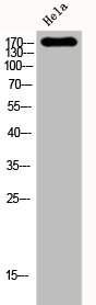
VEGF Receptor 2 antibodyGTX17530
ApplicationsWestern Blot, ImmunoHistoChemistry
TargetKDR
- SizePrice
Product group Antibodies
ApplicationsFlow Cytometry, ImmunoFluorescence, Western Blot, ImmunoCytoChemistry
TargetKDR
- SizePrice
Product group Antibodies
ApplicationsWestern Blot, ELISA, ImmunoHistoChemistry, ImmunoHistoChemistry Frozen
TargetKDR
- SizePrice
![Activation assay analysis of HUVECs pre-treated with 5.0 and 25 microg/ml GTX53462 VEGF Receptor 2 antibody [6B11] and then stimulated with rhVEGF (10ng/ml) for 30min. The phospho-VEGFR2 was detected by detected with IP-Western for P-Tyrosine.](https://www.genetex.com/upload/website/prouct_img/normal/GTX53462/GTX53462_20191119_Activation_w_23060900_230.webp)
Product group Antibodies
References
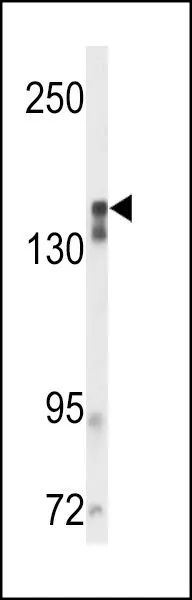
VEGF Receptor 2 antibody [6B11]GTX53462
ApplicationsWestern Blot, Neutralisation/Blocking, Other Application
TargetKDR
- SizePrice
![Activation assay analysis of HUVECs stimulated with 20ng/ml VEGF or treated with 5.0 and 25 microg/ml GTX53464 VEGF Receptor 2 antibody [8A01] (30mins). Phospho-VEGFR2 was detected with IP-Western for P-Tyrosine.](https://www.genetex.com/upload/website/prouct_img/normal/GTX53464/GTX53464_20191119_Activation_w_23060900_578.webp)
Product group Antibodies
VEGF Receptor 2 antibody [8A01]GTX53464
ApplicationsImmunoPrecipitation, Western Blot, Other Application
TargetKDR
- SizePrice
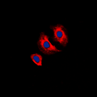
![FACS analysis of HepG2 cells using GTX83308 VEGF Receptor 2 antibody [4B4]. Green : VEGF Receptor 2 Purple : negative control](https://www.genetex.com/upload/website/prouct_img/normal/GTX83308/GTX83308_20170912_FACS_w_23061322_153.webp)
Product group Antibodies
VEGF Receptor 2 antibody [4B4]GTX83308
ApplicationsFlow Cytometry, ImmunoFluorescence, Western Blot, ELISA, ImmunoCytoChemistry
TargetKDR
- SizePrice
Product group Antibodies
VEGF Receptor 2 antibodyGTX80591
ApplicationsWestern Blot, ImmunoHistoChemistry, ImmunoHistoChemistry Paraffin
TargetKDR
- SizePrice
Product group Antibodies
ApplicationsImmunoFluorescence, Western Blot, ImmunoCytoChemistry, ImmunoHistoChemistry, ImmunoHistoChemistry Paraffin
TargetKDR
- SizePrice
Product group Antibodies
ApplicationsImmunoHistoChemistry, ImmunoHistoChemistry Paraffin
TargetKDR
- SizePrice