

Whole cell extract (30 μg) was separated by 10% SDS-PAGE, and the membrane was blotted with Vimentin antibody (GTX629744) diluted at 1:500000. The HRP-conjugated anti-mouse IgG antibody (GTX213111-01) was used to detect the primary antibody, and the signal was developed with Trident ECL plus-Enhanced.
![Various whole cell extracts (30 μg) were separated by 10% SDS-PAGE, and the membrane was blotted with Vimentin antibody [GT812] (GTX629744) diluted at 1:3000.](https://www.genetex.com/upload/website/prouct_img/normal/GTX629744/GTX629744_41421_20161013_WB_M_w_23061202_280.webp "Various whole cell extracts (30 μg) were separated by 10% SDS-PAGE, and the membrane was blotted with Vimentin antibody [GT812] (GTX629744) diluted at 1:3000.")
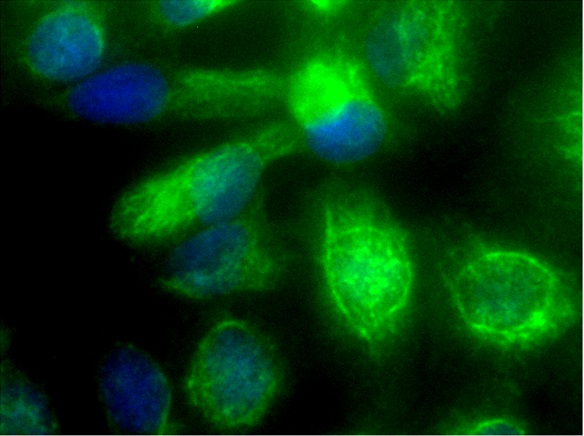
![Vimentin antibody [GT812] detects Vimentin proteins in embryonic mouse brain by immunohistochemical analysis. Sample: Frozen section of embryonic mouse brain (mE18.5). Red: Vimentin antibody [GT812] (GTX629744) diluted at 1:250. Blue: DAPI.](https://www.genetex.com/upload/website/prouct_img/normal/GTX629744/GTX629744_41421_20150703_IHC-Fr_M_w_23061202_937.webp "Vimentin antibody [GT812] detects Vimentin proteins in embryonic mouse brain by immunohistochemical analysis. Sample: Frozen section of embryonic mouse brain (mE18.5). Red: Vimentin antibody [GT812] (GTX629744) diluted at 1:250. Blue: DAPI.")
![Non-transfected (–) and transfected (+) 293T whole cell extracts (30 μg) were separated by 10% SDS-PAGE, and the membrane was blotted with Vimentin antibody [GT812] (GTX629744) diluted at 1:5000.](https://www.genetex.com/upload/website/prouct_img/normal/GTX629744/GTX629744_41421_20160616_WB_shRNA_watermark_w_23061202_797.webp "Non-transfected (–) and transfected (+) 293T whole cell extracts (30 μg) were separated by 10% SDS-PAGE, and the membrane was blotted with Vimentin antibody [GT812] (GTX629744) diluted at 1:5000.")
![Wild-type (WT) and Vimentin knockout (KO) 293T cell extracts (30 μg) were separated by 10% SDS-PAGE, and the membrane was blotted with Vimentin antibody [GT812] (GTX629744) diluted at 1:5000. The HRP-conjugated anti-mouse IgG antibody (GTX213111-01) was used to detect the primary antibody.](https://www.genetex.com/upload/website/prouct_img/normal/GTX629744/GTX629744_41421_20200110_WB_KO_watermark_w_23061202_252.webp "Wild-type (WT) and Vimentin knockout (KO) 293T cell extracts (30 μg) were separated by 10% SDS-PAGE, and the membrane was blotted with Vimentin antibody [GT812] (GTX629744) diluted at 1:5000. The HRP-conjugated anti-mouse IgG antibody (GTX213111-01) was used to detect the primary antibody.")
![Various whole cell extracts (30 μg) were separated by 10% SDS-PAGE, and the membranes were blotted with Vimentin antibody [GT812] (GTX629744) diluted at 1:5000 and competitor's antibody diluted at 1:5000. The HRP-conjugated anti-mouse IgG antibody (GTX213111-01) was used to detect the primary antibody. *The competitor is not affiliated with GeneTex and does not endorse this product.](https://www.genetex.com/upload/website/prouct_img/normal/GTX629744/GTX629744_41421_20200115_WB_competitor_watermark_2_w_23061202_567.webp "Various whole cell extracts (30 μg) were separated by 10% SDS-PAGE, and the membranes were blotted with Vimentin antibody [GT812] (GTX629744) diluted at 1:5000 and competitor's antibody diluted at 1:5000. The HRP-conjugated anti-mouse IgG antibody (GTX213111-01) was used to detect the primary antibody. *The competitor is not affiliated with GeneTex and does not endorse this product.")
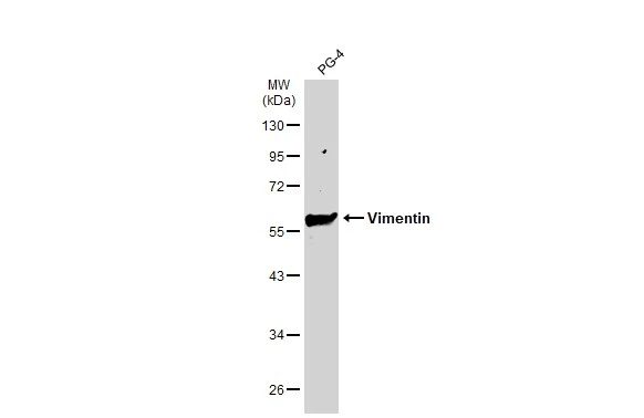
![Whole cell extract (30 μg) was separated by 10% SDS-PAGE, and the membrane was blotted with Vimentin antibody [GT812] (GTX629744) diluted at 1:75000.](https://www.genetex.com/upload/website/prouct_img/normal/GTX629744/GTX629744_41421_20160602_WB_R_w_23061202_533.webp "Whole cell extract (30 μg) was separated by 10% SDS-PAGE, and the membrane was blotted with Vimentin antibody [GT812] (GTX629744) diluted at 1:75000.")
 (GTX213110-01) was diluted at 1:10000 and used to detect the primary antibody.")
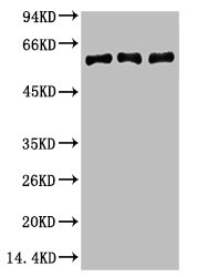
![Various whole cell extracts (30 μg) were separated by 10% SDS-PAGE, and the membrane was blotted with Vimentin antibody [GT812] (GTX629744) diluted at 1:5000.](https://www.genetex.com/upload/website/prouct_img/normal/GTX629744/GTX629744_41421_20160505_WB_w_23061202_705.webp "Various whole cell extracts (30 μg) were separated by 10% SDS-PAGE, and the membrane was blotted with Vimentin antibody [GT812] (GTX629744) diluted at 1:5000.")
![Non-transfected (–) and transfected (+) 293T whole cell extracts (10 μg) were separated by 10% SDS-PAGE, and the membrane was blotted with Vimentin antibody [GT812] (GTX629744) diluted at 1:10000. The HRP-conjugated anti-mouse IgG antibody (GTX213111-01) was used to detect the primary antibody.](https://www.genetex.com/upload/website/prouct_img/normal/GTX629744/GTX629744_41421_20200925_WB_B_w_23061202_761.webp "Non-transfected (–) and transfected (+) 293T whole cell extracts (10 μg) were separated by 10% SDS-PAGE, and the membrane was blotted with Vimentin antibody [GT812] (GTX629744) diluted at 1:10000. The HRP-conjugated anti-mouse IgG antibody (GTX213111-01) was used to detect the primary antibody.")
Whole cell extract (30 μg) was separated by 10% SDS-PAGE, and the membrane was blotted with Vimentin antibody (GTX629744) diluted at 1:500000. The HRP-conjugated anti-mouse IgG antibody (GTX213111-01) was used to detect the primary antibody, and the signal was developed with Trident ECL plus-Enhanced.
Vimentin antibody [GT812]
GTX629744
ApplicationsFlow Cytometry, ImmunoFluorescence, ImmunoPrecipitation, Western Blot, ELISA, ImmunoCytoChemistry, ImmunoHistoChemistry, ImmunoHistoChemistry Frozen, ImmunoHistoChemistry Paraffin
Product group Antibodies
ReactivityFeline, Human, Mouse, Rat
TargetVIM
Overview
- SupplierGeneTex
- Product NameVimentin antibody [GT812]
- Delivery Days Customer9
- Application Supplier NoteWB: 1:500-1:75000. IHC-P: 1:100-1:1000. IHC-Fr: 1:100-1:1000. *Optimal dilutions/concentrations should be determined by the researcher.Not tested in other applications.
- ApplicationsFlow Cytometry, ImmunoFluorescence, ImmunoPrecipitation, Western Blot, ELISA, ImmunoCytoChemistry, ImmunoHistoChemistry, ImmunoHistoChemistry Frozen, ImmunoHistoChemistry Paraffin
- CertificationResearch Use Only
- ClonalityMonoclonal
- Clone IDGT812
- Concentration1 mg/ml
- ConjugateUnconjugated
- Gene ID7431
- Target nameVIM
- Target descriptionvimentin
- Target synonymsvimentin, epididymis secretory sperm binding protein
- HostMouse
- IsotypeIgG2b
- Protein IDP08670
- Protein NameVimentin
- Scientific DescriptionAlong with the microfilaments (actins) and microtubules (tubulins), the intermediate filaments represent a third class of well-characterized cytoskeletal elements. The subunits display a tissue-specific pattern of expression. Desmin (MIM 125660) is the subunit specific for muscle and vimentin the subunit specific for mesenchymal tissue.[supplied by OMIM]
- ReactivityFeline, Human, Mouse, Rat
- Storage Instruction-20°C or -80°C,2°C to 8°C
- UNSPSC12352203
References
- Hurtado-Alvarado G, Soto-Tinoco E, Santacruz-Martínez E, et al. Suprachiasmatic nucleus promotes hyperglycemia induced by sleep delay. Curr Biol. 2023,33(20):4343-4352.e4. doi: 10.1016/j.cub.2023.08.071Read this paper
- Yang KS, O'Shea A, Zelga P, et al. Extracellular vesicle analysis of plasma allows differential diagnosis of atypical pancreatic serous cystadenoma. Sci Rep. 2023,13(1):10969. doi: 10.1038/s41598-023-37966-5Read this paper
- Wanandi SI, Hilbertina N, Siregar NC, et al. Cancer-associated fibroblast (CAF) secretomes-induced epithelial-mesenchymal transition on HT-29 colorectal carcinoma cells associated with hepatocyte growth factor (HGF) signalling. J Pak Med Assoc. 2021,71(Suppl 2)(2):S18-S24.Read this paper
- Liu L, Meng T, Zheng X, et al. Transgelin 2 Promotes Paclitaxel Resistance, Migration, and Invasion of Breast Cancer by Directly Interacting with PTEN and Activating PI3K/Akt/GSK-3β Pathway. Mol Cancer Ther. 2019,18(12):2457-2468. doi: 10.1158/1535-7163.MCT-19-0261Read this paper
- Lee SW, Chen YW, Kuan EC, et al. Dual-function nanostructured platform for isolation of nasopharyngeal carcinoma circulating tumor cells and EBV DNA detection. Biosens Bioelectron. 2019,142:111509. doi: 10.1016/j.bios.2019.111509Read this paper
- Sikorski K, Mehta A, Inngjerdingen M, et al. A high-throughput pipeline for validation of antibodies. Nat Methods. 2018,15(11):909-912. doi: 10.1038/s41592-018-0179-8Read this paper
- Liu MC, Chen WH, Chiou CS, et al. Inhibition of chronic prostate inflammation by hyaluronic acid through an immortalized human prostate stromal cell line model. PLoS One. 2017,12(5):e0178152. doi: 10.1371/journal.pone.0178152Read this paper
- Jung H, Kim B, Moon BI, et al. Cytokeratin 18 is necessary for initiation of TGF-β1-induced epithelial-mesenchymal transition in breast epithelial cells. Mol Cell Biochem. 2016,423(1-2):21-28.Read this paper
- Rakhila H, Al-Akoum M, Doillon C, et al. Augmented Angiogenic Factors Expression via FP Signaling Pathways in Peritoneal Endometriosis. J Clin Endocrinol Metab. 2016,101(12):4752-4763.Read this paper
Datasheet
Related products
Product group Antibodies

Anti-Vimentin [1433]AB01640-1.9-C
ApplicationsImmunoFluorescence
ReactivityCanine, Human, Rat
TargetVIM
- SizePrice

Product group Antibodies
Anti-VIM AntibodyAMAB90516
ApplicationsWestern Blot, ImmunoHistoChemistry
ReactivityHuman
TargetVIM
- SizePrice
Product group Antibodies
Vim Polyclonal AntibodyCAC07060
ApplicationsImmunoFluorescence, Western Blot, ELISA, ImmunoHistoChemistry
TargetVIM
- SizePrice
Product group Antibodies
References
Vimentin Polyclonal AntibodyBS-0756R
ApplicationsFlow Cytometry, ImmunoFluorescence, Western Blot, ELISA, ImmunoCytoChemistry, ImmunoHistoChemistry, ImmunoHistoChemistry Frozen, ImmunoHistoChemistry Paraffin
ReactivityBovine, Chicken, Goat, Human, Mouse, Porcine, Rat
TargetVIM
- SizePrice
Product group Antibodies
VIM Monoclonal AntibodyCSB-MA000319
ApplicationsWestern Blot, ELISA
ReactivityHuman, Mouse, Rat
TargetVIM
- SizePrice
Product group Antibodies
Vimentin antibody [SRL33]GTX01972
ApplicationsImmunoHistoChemistry, ImmunoHistoChemistry Paraffin
ReactivityHuman
TargetVIM
- SizePrice
Product group Antibodies
Vimentin antibodyGTX82958
ApplicationsImmunoHistoChemistry
ReactivityHuman
TargetVIM
- SizePrice