

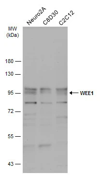
Various whole cell extracts (30 μg) were separated by 7.5% SDS-PAGE, and the membrane was blotted with WEE1 antibody [N3C2], Internal (GTX111392) diluted at 1:500. The HRP-conjugated anti-rabbit IgG antibody (GTX213110-01) was used to detect the primary antibody.
![Various whole cell extracts (30 μg) were separated by 7.5% SDS-PAGE, and the membranes were blotted with WEE1 antibody [N3C2], Internal (GTX111392) diluted at 1:1000 and competitor's antibody (CST#13084) diluted at 1:1000. The HRP-conjugated anti-rabbit IgG antibody (GTX213110-01) was used to detect the primary antibody.](https://www.genetex.com/upload/website/prouct_img/normal/GTX111392/GTX111392_42795_20170728_WB_competitor_watermark_w_23060500_623.webp "Various whole cell extracts (30 μg) were separated by 7.5% SDS-PAGE, and the membranes were blotted with WEE1 antibody [N3C2], Internal (GTX111392) diluted at 1:1000 and competitor's antibody (CST#13084) diluted at 1:1000. The HRP-conjugated anti-rabbit IgG antibody (GTX213110-01) was used to detect the primary antibody.")
 antibody at 1:100 dilution.
Antigen Retrieval: Trilogy? (EDTA based, pH 8.0) buffer, 15min")
![Various whole cell extracts (30 μg) were separated by 7.5% SDS-PAGE, and the membrane was blotted with WEE1 antibody [N3C2], Internal (GTX111392) diluted at 1:1000. The HRP-conjugated anti-rabbit IgG antibody (GTX213110-01) was used to detect the primary antibody.](https://www.genetex.com/upload/website/prouct_img/normal/GTX111392/GTX111392_45238_20231124_WB_25061003_773.webp "Various whole cell extracts (30 μg) were separated by 7.5% SDS-PAGE, and the membrane was blotted with WEE1 antibody [N3C2], Internal (GTX111392) diluted at 1:1000. The HRP-conjugated anti-rabbit IgG antibody (GTX213110-01) was used to detect the primary antibody.")
![Various whole cell extracts (30 μg) were separated by 7.5% SDS-PAGE, and the membrane was blotted with WEE1 antibody [N3C2], Internal (GTX111392) diluted at 1:1000. The HRP-conjugated anti-rabbit IgG antibody (GTX213110-01) was used to detect the primary antibody. Corresponding RNA expression data for the same cell lines are based on Human Protein Atlas program.](https://www.genetex.com/upload/website/prouct_img/normal/GTX111392/GTX111392_45238_20250124_WB_TPM_watermark_25061003_899.webp "Various whole cell extracts (30 μg) were separated by 7.5% SDS-PAGE, and the membrane was blotted with WEE1 antibody [N3C2], Internal (GTX111392) diluted at 1:1000. The HRP-conjugated anti-rabbit IgG antibody (GTX213110-01) was used to detect the primary antibody. Corresponding RNA expression data for the same cell lines are based on Human Protein Atlas program.")
![Mouse tissue extract (50 μg) was separated by 7.5% SDS-PAGE, and the membrane was blotted with WEE1 antibody [N3C2], Internal (GTX111392) diluted at 1:1000. The HRP-conjugated anti-rabbit IgG antibody (GTX213110-01) was used to detect the primary antibody.](https://www.genetex.com/upload/website/prouct_img/normal/GTX111392/GTX111392_45238_20231124_WB_M_spleen_25061003_134.webp "Mouse tissue extract (50 μg) was separated by 7.5% SDS-PAGE, and the membrane was blotted with WEE1 antibody [N3C2], Internal (GTX111392) diluted at 1:1000. The HRP-conjugated anti-rabbit IgG antibody (GTX213110-01) was used to detect the primary antibody.")
Various whole cell extracts (30 μg) were separated by 7.5% SDS-PAGE, and the membrane was blotted with WEE1 antibody [N3C2], Internal (GTX111392) diluted at 1:500. The HRP-conjugated anti-rabbit IgG antibody (GTX213110-01) was used to detect the primary antibody.
WEE1 antibody [N3C2], Internal
GTX111392
ApplicationsWestern Blot, ImmunoHistoChemistry, ImmunoHistoChemistry Paraffin
Product group Antibodies
ReactivityHuman, Mouse
TargetWEE1
Overview
- SupplierGeneTex
- Product NameWEE1 antibody [N3C2], Internal
- Delivery Days Customer9
- Application Supplier NoteWB: 1:500-1:3000. IHC-P: 1:100-1:1000. *Optimal dilutions/concentrations should be determined by the researcher.Not tested in other applications.
- ApplicationsWestern Blot, ImmunoHistoChemistry, ImmunoHistoChemistry Paraffin
- CertificationResearch Use Only
- ClonalityPolyclonal
- Concentration1.45 mg/ml
- ConjugateUnconjugated
- Gene ID7465
- Target nameWEE1
- Target descriptionWEE1 G2 checkpoint kinase
- Target synonymsWEE1A, WEE1hu, wee1-like protein kinase, WEE1 homolog, WEE1+ homolog, protein kinase, wee1A kinase
- HostRabbit
- IsotypeIgG
- Protein IDP30291
- Protein NameWee1-like protein kinase
- Scientific DescriptionThis gene encodes a nuclear protein, which is a tyrosine kinase belonging to the Ser/Thr family of protein kinases. This protein catalyzes the inhibitory tyrosine phosphorylation of CDC2/cyclin B kinase, and appears to coordinate the transition between DNA replication and mitosis by protecting the nucleus from cytoplasmically activated CDC2 kinase. [provided by RefSeq]
- ReactivityHuman, Mouse
- Storage Instruction-20°C or -80°C,2°C to 8°C
- UNSPSC41116161
Datasheet
Related products
Product group Antibodies
Anti-WEE1 AntibodyA97865
ApplicationsWestern Blot, ELISA
ReactivityHuman, Mouse, Rat
- SizePrice
Product group Antibodies
Anti-Wee1 (S123) Antibody102-25272
ApplicationsWestern Blot
TargetWEE1
- SizePrice

Product group Antibodies
Anti-WEE1 Antibody Picoband(r)A01319-1-CARRIER-FREE
ApplicationsFlow Cytometry, ImmunoFluorescence, Western Blot, ImmunoCytoChemistry, ImmunoHistoChemistry
ReactivityHuman, Mouse, Rat
TargetWEE1
- SizePrice

Product group Antibodies

WEE1 AntibodyLS-C834919
ApplicationsELISA, ImmunoHistoChemistry
ReactivityHuman, Mouse, Rat
TargetWEE1
- SizePrice
Product group Antibodies
WEE1 Polyclonal Antibody, AbBy Fluor-350 ConjugatedBS-23526R-BF350
ApplicationsImmunoFluorescence, Western Blot, ImmunoCytoChemistry, ImmunoHistoChemistry, ImmunoHistoChemistry Frozen, ImmunoHistoChemistry Paraffin
ReactivityHuman, Mouse, Rat
TargetWEE1
- SizePrice
Product group Antibodies
Phospho-WEE1 (S642) AntibodyCSB-PA060083
ApplicationsWestern Blot, ELISA
ReactivityHuman, Mouse, Rat
TargetWEE1
- SizePrice

Product group Antibodies
Anti-WEE1 AntibodyHPA068845
ApplicationsWestern Blot
ReactivityHuman
TargetWEE1
- SizePrice
Product group Antibodies
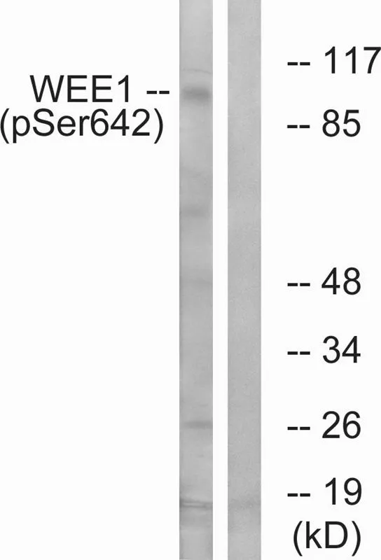
WEE1 (phospho Ser642) antibodyGTX55324
ApplicationsWestern Blot
ReactivityHuman
TargetWEE1
- SizePrice
Product group Antibodies
Anti-WEE1Y058162
ApplicationsWestern Blot, ELISA
ReactivityHuman, Rat
- SizePrice