

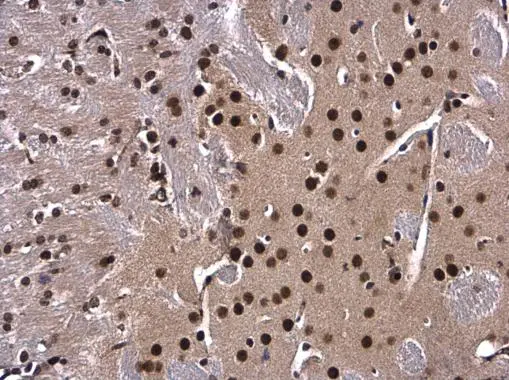
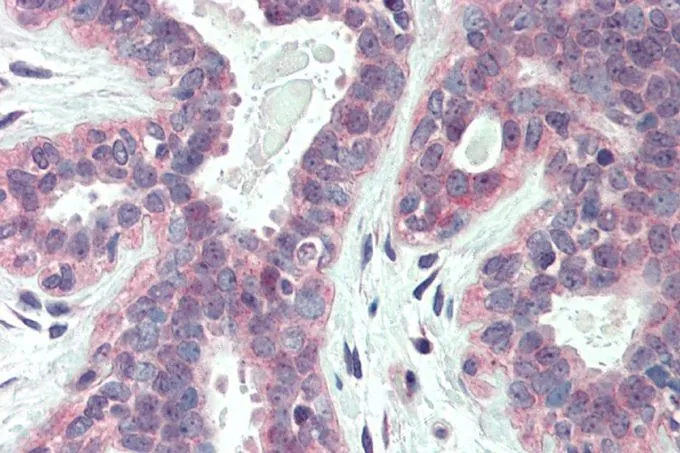
XBP1 antibody [N3C3] detects XBP1 protein at nucleus in mouse brain by immunohistochemical analysis. Sample: Paraffin-embedded mouse brain. XBP1 antibody [N3C3] (GTX102229) diluted at 1:500.
Antigen Retrieval: Citrate buffer, pH 6.0, 15 min
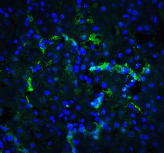
![XBP1 antibody [N3C3] detects XBP1 protein at nucleus by immunofluorescent analysis. Sample: HepG2 cells were fixed in 4% paraformaldehyde at RT for 15 min. Green: XBP1 stained by XBP1 antibody [N3C3] (GTX102229) diluted at 1:500. Blue: Fluoroshield with DAPI (GTX30920).](https://www.genetex.com/upload/website/prouct_img/normal/GTX102229/GTX102229_44286_20220121_ICC_IF_w_23060100_733.webp "XBP1 antibody [N3C3] detects XBP1 protein at nucleus by immunofluorescent analysis. Sample: HepG2 cells were fixed in 4% paraformaldehyde at RT for 15 min. Green: XBP1 stained by XBP1 antibody [N3C3] (GTX102229) diluted at 1:500. Blue: Fluoroshield with DAPI (GTX30920).")
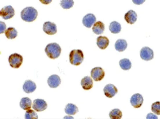
![XBP1 antibody [N3C3] detects XBP1 protein at cytoplasm and nucleus by immunohistochemical analysis. Sample: Paraffin-embedded human breast carcinoma. XBP1 stained by XBP1 antibody [N3C3] (GTX102229) diluted at 1:1000. Antigen Retrieval: Citrate buffer, pH 6.0, 15 min](https://www.genetex.com/upload/website/prouct_img/normal/GTX102229/GTX102229_43901_20200529_IHC-P_w_23060100_523.webp "XBP1 antibody [N3C3] detects XBP1 protein at cytoplasm and nucleus by immunohistochemical analysis. Sample: Paraffin-embedded human breast carcinoma. XBP1 stained by XBP1 antibody [N3C3] (GTX102229) diluted at 1:1000. Antigen Retrieval: Citrate buffer, pH 6.0, 15 min")
![XBP1 antibody [N3C3] detects XBP1 protein at nucleus in rat ovary by immunohistochemical analysis. Sample: Paraffin-embedded rat ovary. XBP1 antibody [N3C3] (GTX102229) diluted at 1:500.
Antigen Retrieval: Citrate buffer, pH 6.0, 15 min](https://www.genetex.com/upload/website/prouct_img/normal/GTX102229/GTX102229_40079_20151130_IHC-P_R_w_23060100_386.webp "XBP1 antibody [N3C3] detects XBP1 protein at nucleus in rat ovary by immunohistochemical analysis. Sample: Paraffin-embedded rat ovary. XBP1 antibody [N3C3] (GTX102229) diluted at 1:500.
Antigen Retrieval: Citrate buffer, pH 6.0, 15 min")
 antibody at 1:500 dilution.
Antigen Retrieval: Citrate buffer, pH 6.0, 15 min")
![Untreated (–) and treated (+) HepG2 whole cell extracts (30 μg) were separated by 10% SDS-PAGE, and the membrane was blotted with XBP1 antibody [N3C3] (GTX102229) diluted at 1:1000. The HRP-conjugated anti-rabbit IgG antibody (GTX213110-01) was used to detect the primary antibody.](https://www.genetex.com/upload/website/prouct_img/normal/GTX102229/GTX102229_45490_20240816_WB_treatment_Thapsigargin_24082301_561.webp "Untreated (–) and treated (+) HepG2 whole cell extracts (30 μg) were separated by 10% SDS-PAGE, and the membrane was blotted with XBP1 antibody [N3C3] (GTX102229) diluted at 1:1000. The HRP-conjugated anti-rabbit IgG antibody (GTX213110-01) was used to detect the primary antibody.")
XBP1 antibody [N3C3] detects XBP1 protein at nucleus in mouse brain by immunohistochemical analysis. Sample: Paraffin-embedded mouse brain. XBP1 antibody [N3C3] (GTX102229) diluted at 1:500.
Antigen Retrieval: Citrate buffer, pH 6.0, 15 min
XBP1 antibody [N3C3]
GTX102229
ApplicationsImmunoFluorescence, Western Blot, ImmunoCytoChemistry, ImmunoHistoChemistry, ImmunoHistoChemistry Paraffin
Product group Antibodies
ReactivityHuman, Mouse, Rat
TargetXBP1
Overview
- SupplierGeneTex
- Product NameXBP1 antibody [N3C3]
- Delivery Days Customer9
- Application Supplier NoteWB: 1:500-1:3000. ICC/IF: 1:100-1:1000. IHC-P: 1:100-1:1000. *Optimal dilutions/concentrations should be determined by the researcher.Not tested in other applications.
- ApplicationsImmunoFluorescence, Western Blot, ImmunoCytoChemistry, ImmunoHistoChemistry, ImmunoHistoChemistry Paraffin
- CertificationResearch Use Only
- ClonalityPolyclonal
- Concentration0.52 mg/ml
- ConjugateUnconjugated
- Gene ID7494
- Target nameXBP1
- Target descriptionX-box binding protein 1
- Target synonymsTREB-5, TREB5, XBP-1, XBP2, X-box-binding protein 1, tax-responsive element-binding protein 5
- HostRabbit
- IsotypeIgG
- Protein IDP17861
- Protein NameX-box-binding protein 1
- Scientific DescriptionThis gene encodes a transcription factor that regulates MHC class II genes by binding to a promoter element referred to as an X box. This gene product is a bZIP protein, which was also identified as a cellular transcription factor that binds to an enhancer in the promoter of the T cell leukemia virus type 1 promoter. It may increase expression of viral proteins by acting as the DNA binding partner of a viral transactivator. It has been found that upon accumulation of unfolded proteins in the endoplasmic reticulum (ER), the mRNA of this gene is processed to an active form by an unconventional splicing mechanism that is mediated by the endonuclease inositol-requiring enzyme 1 (IRE1). The resulting loss of 26 nt from the spliced mRNA causes a frame-shift and an isoform XBP1(S), which is the functionally active transcription factor. The isoform encoded by the unspliced mRNA, XBP1(U), is constitutively expressed, and thought to function as a negative feedback regulator of XBP1(S), which shuts off transcription of target genes during the recovery phase of ER stress. A pseudogene of XBP1 has been identified and localized to chromosome 5. [provided by RefSeq]
- ReactivityHuman, Mouse, Rat
- Storage Instruction-20°C or -80°C,2°C to 8°C
- UNSPSC41116161
Datasheet
Related products
Product group Antibodies
Anti-XBP1 Antibody144-01731
ApplicationsImmunoFluorescence, Western Blot
ReactivityHuman, Mouse, Rat
TargetXBP1
- SizePrice
Product group Antibodies
Anti-XBP1 AntibodyA13620
ApplicationsImmunoFluorescence, Western Blot, ImmunoCytoChemistry
ReactivityHuman, Mouse, Rat
- SizePrice
Product group Antibodies

Goat anti-XBP1 / TREB5EB08557
ApplicationsImmunoFluorescence, Western Blot, ELISA, ImmunoHistoChemistry
ReactivityCanine, Human, Mouse, Rat
TargetXBP1
- SizePrice
Product group Antibodies
XBP1 AntibodyCSB-PA027191
ApplicationsELISA, ImmunoHistoChemistry
ReactivityHuman
TargetXBP1
- SizePrice
Product group Antibodies
References
XBP1 Polyclonal AntibodyBS-1668R
ApplicationsFlow Cytometry, ImmunoFluorescence, Western Blot, ELISA, ImmunoCytoChemistry, ImmunoHistoChemistry, ImmunoHistoChemistry Frozen, ImmunoHistoChemistry Paraffin
ReactivityBovine, Chicken, Human, Mouse, Rat
TargetXBP1
- SizePrice
Product group Antibodies

XBP1 AntibodyLS-C401118
ApplicationsELISA, ImmunoHistoChemistry
ReactivityHuman
TargetXBP1
- SizePrice
Product group Antibodies
XBP1 antibodyGTX31292
ApplicationsImmunoFluorescence, Western Blot, ELISA, ImmunoCytoChemistry
ReactivityHuman, Mouse, Rat
TargetXBP1
- SizePrice
Product group Antibodies
XBP1 antibodyGTX31293
ApplicationsImmunoFluorescence, Western Blot, ELISA, ImmunoCytoChemistry, ImmunoHistoChemistry, ImmunoHistoChemistry Paraffin
ReactivityHuman, Mouse, Rat
TargetXBP1
- SizePrice
![WB analysis of XBP1(AA: 1-160)-hIgGFc transfected HEK293 cell lysate using GTX83279 XBP1 antibody [1C4].](https://www.genetex.com/upload/website/prouct_img/normal/GTX83279/GTX83279_20170912_WB_w_23061322_946.webp)
Product group Antibodies
XBP1 antibody [1C4]GTX83279
ApplicationsWestern Blot, ELISA
ReactivityHuman
TargetXBP1
- SizePrice
Product group Antibodies
XBP1 antibody, InternalGTX88727
ApplicationsWestern Blot, ImmunoHistoChemistry, ImmunoHistoChemistry Paraffin
ReactivityHuman
TargetXBP1
- SizePrice