

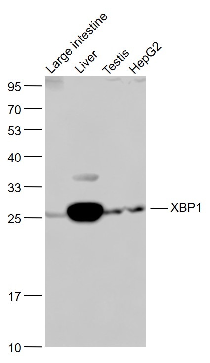
Lane 1: Mouse Large intestine lysates; Lane 2: Mouse Liver lysates; Lane 3: Mouse Testis lysates; Lane 4: HepG2 cell lysates probed with XBP1 Polyclonal Antibody, Unconjugated (bs-1668R) at 1:1000 dilution and 4˚C overnight incubation. Followed by conjugated secondary antibody incubation at 1:20000 for 60 min at 37˚C.
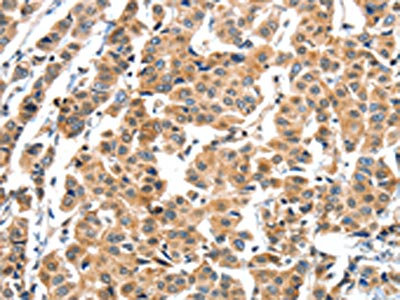
 at 37°C for 20 min; Antibody incubation with (XBP1) polyclonal Antibody, Unconjugated (bs-1668R) 1:100, 90 minutes at 37°C; followed by a conjugated Goat Anti-Rabbit IgG antibody at 37°C for 90 minutes, DAPI (blue, C02-04002) was used to stain the cell nuclei.")
 at 1:1000 dilution and 4°C overnight incubation. Followed by conjugated secondary antibody incubation at 1:20000 for 60 min at 37˚C.")
 at 1:500 dilution and 4°C overnight incubation. Followed by conjugated secondary antibody incubation at 1:20000 for 60 min at 37˚C.")
 followed by conjugation to the secondary antibody and DAB staining")
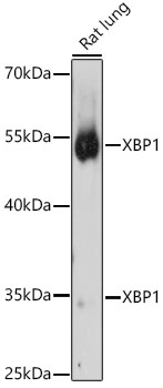
 at 1:200 overnight at 4˚C. Followed by conjugation to secondary antibody (bs-0295G-HRP) at 1:3000 for 90 min at 37˚C. Predicted band 29kD. Observed band size: 29kD")
Lane 1: Mouse Large intestine lysates; Lane 2: Mouse Liver lysates; Lane 3: Mouse Testis lysates; Lane 4: HepG2 cell lysates probed with XBP1 Polyclonal Antibody, Unconjugated (bs-1668R) at 1:1000 dilution and 4˚C overnight incubation. Followed by conjugated secondary antibody incubation at 1:20000 for 60 min at 37˚C.
XBP1 Polyclonal Antibody
BS-1668R
ApplicationsFlow Cytometry, ImmunoFluorescence, Western Blot, ELISA, ImmunoCytoChemistry, ImmunoHistoChemistry, ImmunoHistoChemistry Frozen, ImmunoHistoChemistry Paraffin
Product group Antibodies
ReactivityBovine, Chicken, Human, Mouse, Rat
TargetXBP1
Overview
- SupplierBioss
- Product NameXBP1 Polyclonal Antibody
- Delivery Days Customer16
- ApplicationsFlow Cytometry, ImmunoFluorescence, Western Blot, ELISA, ImmunoCytoChemistry, ImmunoHistoChemistry, ImmunoHistoChemistry Frozen, ImmunoHistoChemistry Paraffin
- Applications SupplierWB(1:300-5000), ELISA(1:500-1000), FCM(1:20-100), IHC-P(1:200-400), IHC-F(1:100-500), IF(IHC-P)(1:50-200), IF(IHC-F)(1:50-200), IF(ICC)(1:50-200)
- CertificationResearch Use Only
- ClonalityPolyclonal
- Concentration1 ug/ul
- ConjugateUnconjugated
- Gene ID7494
- Target nameXBP1
- Target descriptionX-box binding protein 1
- Target synonymsTREB-5, TREB5, XBP-1, XBP2, X-box-binding protein 1, tax-responsive element-binding protein 5
- HostRabbit
- IsotypeIgG
- Protein IDP17861
- Protein NameX-box-binding protein 1
- ReactivityBovine, Chicken, Human, Mouse, Rat
- Storage Instruction-20°C
- UNSPSC41116161
References
- Radioprotective effect of Hohenbuehelia serotina polysaccharides through mediation of ER apoptosis pathway in vivo. Wang L et al., 2019 Apr 15, Int J Biol MacromolRead this paper
- Recombinant human maspin inhibits high glucose-induced oxidative stress and angiogenesis of human retinal microvascular endothelial cells via PI3K/AKT pathway. Qiu F et al., 2018 Sep, Mol Cell BiochemRead this paper
- Silver nanoparticles induce SH-SY5Y cell apoptosis via endoplasmic reticulum- and mitochondrial pathways that lengthen endoplasmic reticulum-mitochondria contact sites and alter inositol-3-phosphate receptor function. Li L et al., 2018 Mar 15, Toxicol LettRead this paper
Datasheet
Related products
Product group Antibodies
Anti-XBP1 Antibody144-01731
ApplicationsImmunoFluorescence, Western Blot
ReactivityHuman, Mouse, Rat
TargetXBP1
- SizePrice
Product group Antibodies
Anti-XBP1 AntibodyA13620
ApplicationsImmunoFluorescence, Western Blot, ImmunoCytoChemistry
ReactivityHuman, Mouse, Rat
- SizePrice
Product group Antibodies

Goat anti-XBP1 / TREB5EB08557
ApplicationsImmunoFluorescence, Western Blot, ELISA, ImmunoHistoChemistry
ReactivityCanine, Human, Mouse, Rat
TargetXBP1
- SizePrice
Product group Antibodies
XBP1 AntibodyCSB-PA027191
ApplicationsELISA, ImmunoHistoChemistry
ReactivityHuman
TargetXBP1
- SizePrice
Product group Antibodies

XBP1 AntibodyLS-C401118
ApplicationsELISA, ImmunoHistoChemistry
ReactivityHuman
TargetXBP1
- SizePrice
![XBP1 antibody [N3C3] detects XBP1 protein at nucleus in mouse brain by immunohistochemical analysis. Sample: Paraffin-embedded mouse brain. XBP1 antibody [N3C3] (GTX102229) diluted at 1:500.
Antigen Retrieval: Citrate buffer, pH 6.0, 15 min](https://www.genetex.com/upload/website/prouct_img/normal/GTX102229/GTX102229_40079_20151130_IHC-P_M_w_23060100_471.webp)
Product group Antibodies
XBP1 antibody [N3C3]GTX102229
ApplicationsImmunoFluorescence, Western Blot, ImmunoCytoChemistry, ImmunoHistoChemistry, ImmunoHistoChemistry Paraffin
ReactivityHuman, Mouse, Rat
TargetXBP1
- SizePrice

Product group Antibodies
Anti-XBP1 Antibody Picoband(r)PB9463-CARRIER-FREE
ApplicationsFlow Cytometry, ImmunoFluorescence, Western Blot, ImmunoCytoChemistry, ImmunoHistoChemistry
ReactivityHamster, Human, Mouse, Rat
TargetXBP1
- SizePrice