

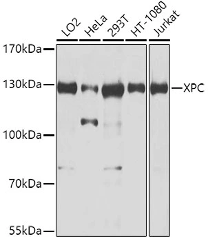
WB analysis of various sample lysates using GTX55845 XPC antibody. The signal was developed with ECL plus-Enhanced. Dilution : 1:1000 Loading : 25μg per lane


WB analysis of various sample lysates using GTX55845 XPC antibody. The signal was developed with ECL plus-Enhanced. Dilution : 1:1000 Loading : 25μg per lane
XPC antibody
GTX55845
ApplicationsImmunoPrecipitation, Western Blot, ImmunoHistoChemistry, ImmunoHistoChemistry Paraffin
Product group Antibodies
ReactivityHuman
TargetXPC
Overview
- SupplierGeneTex
- Product NameXPC antibody
- Delivery Days Customer9
- Application Supplier NoteWB: 1:500 - 1:2000. IHC-P: 1:50 - 1:100. IP: 1:50 - 1:100. *Optimal dilutions/concentrations should be determined by the researcher.Not tested in other applications.
- ApplicationsImmunoPrecipitation, Western Blot, ImmunoHistoChemistry, ImmunoHistoChemistry Paraffin
- CertificationResearch Use Only
- ClonalityPolyclonal
- ConjugateUnconjugated
- Gene ID7508
- Target nameXPC
- Target descriptionXPC complex subunit, DNA damage recognition and repair factor
- Target synonymsRAD4, XP3, XPCC, p125, DNA repair protein complementing XP-C cells, mutant xeroderma pigmentosum group C, xeroderma pigmentosum, complementation group C
- HostRabbit
- IsotypeIgG
- Protein IDQ01831
- Protein NameDNA repair protein complementing XP-C cells
- Scientific DescriptionThe protein encoded by this gene is a key component of the XPC complex, which plays an important role in the early steps of global genome nucleotide excision repair (NER). The encoded protein is important for damage sensing and DNA binding, and shows a preference for single-stranded DNA. Mutations in this gene or some other NER components can result in Xeroderma pigmentosum, a rare autosomal recessive disorder characterized by increased sensitivity to sunlight with the development of carcinomas at an early age. Alternatively spliced transcript variants have been found for this gene. [provided by RefSeq, Aug 2017]
- ReactivityHuman
- Storage Instruction-20°C or -80°C,2°C to 8°C
- UNSPSC12352203
Datasheet
Related products
Product group Antibodies
Anti-XPC Antibody144-08354
ApplicationsImmunoPrecipitation, Western Blot, ImmunoHistoChemistry
ReactivityHuman
TargetXPC
- SizePrice
Product group Antibodies
XPC Polyclonal AntibodyBS-25269R
ApplicationsWestern Blot, ELISA
ReactivityBovine, Equine, Human, Mouse, Porcine, Rat
TargetXPC
- SizePrice

Product group Antibodies
Anti-XPC AntibodyA10400
ApplicationsImmunoPrecipitation, Western Blot, ImmunoHistoChemistry
ReactivityHuman
- SizePrice
![XPC antibody [HL2892] detects XPC protein by immunofluorescent analysis. Sample: MCF-7 cells were fixed in ice-cold MeOH for 5 min. Green: XPC stained by XPC antibody [HL2892] (GTX640229) diluted at 1:500. Red: alpha Tubulin, a cytoskeleton marker, stained by alpha Tubulin antibody [GT114] (GTX628802) diluted at 1:1000.](https://www.genetex.com/upload/website/prouct_img/normal/GTX640229/GTX640229_T-45376_20240607_ICC_IF_24062501_773.webp)
Product group Antibodies
XPC antibody [HL2892]GTX640229
ApplicationsImmunoFluorescence, Western Blot, ImmunoCytoChemistry
ReactivityHuman
TargetXPC
- SizePrice
![XPC antibody [HL2894] detects XPC protein by immunohistochemical analysis. Sample: Paraffin-embedded mouse kidney. XPC stained by XPC antibody [HL2894] (GTX640231) diluted at 1:100. Antigen Retrieval: Citrate buffer, pH 6.0, 15 min](https://www.genetex.com/upload/website/prouct_img/normal/GTX640231/GTX640231_T-45376_20240606_IHC-P_M_24061802_534.webp)
Product group Antibodies
XPC antibody [HL2894]GTX640231
ApplicationsWestern Blot, ImmunoHistoChemistry, ImmunoHistoChemistry Paraffin
ReactivityHuman, Mouse, Rat
TargetXPC
- SizePrice
Product group Antibodies

XPC AntibodyLS-C832647
ApplicationsELISA, ImmunoHistoChemistry
ReactivityHuman
TargetXPC
- SizePrice

Product group Antibodies
Anti-XPC AntibodyHPA035707
ApplicationsWestern Blot, ImmunoCytoChemistry, ImmunoHistoChemistry
ReactivityHuman
TargetXPC
- SizePrice
Product group Antibodies
XPC AntibodyCSB-PA026217LA01HU
ApplicationsELISA
ReactivityHuman
TargetXPC
- SizePrice