

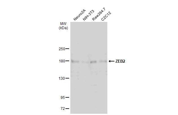
Various whole cell extracts (30 μg) were separated by 5% SDS-PAGE, and the membrane was blotted with ZEB2 antibody (GTX129243) diluted at 1:500. The HRP-conjugated anti-rabbit IgG antibody (GTX213110-01) was used to detect the primary antibody, and the signal was developed with Trident ECL plus-Enhanced.
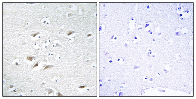
 diluted at 1:1000. Antigen Retrieval: Citrate buffer, pH 6.0, 15 min")
 was separated by 5% SDS-PAGE, and the membrane was blotted with ZEB2 antibody (GTX129243) diluted at 1:500. The HRP-conjugated anti-rabbit IgG antibody (GTX213110-01) was used to detect the primary antibody.")
 were separated by 5% SDS-PAGE, and the membrane was blotted with ZEB2 antibody (GTX129243) diluted at 1:1000. The HRP-conjugated anti-rabbit IgG antibody (GTX213110-01) was used to detect the primary antibody, and the signal was developed with Trident ECL plus-Enhanced.")
![ZEB2 antibody detects ZEB2 protein at nucleus by immunofluorescent analysis. Sample: U87-MG cells were fixed in 4% paraformaldehyde at RT for 15 min. Green: ZEB2 stained by ZEB2 antibody (GTX129243) diluted at 1:500. Red: alpha Tubulin, a cytoskeleton marker, stained by alpha Tubulin antibody [GT114] (GTX628802) diluted at 1:1000.](https://www.genetex.com/upload/website/prouct_img/normal/GTX129243/GTX129243_43565_20220311_ICC_IF_w_23060523_477.webp "ZEB2 antibody detects ZEB2 protein at nucleus by immunofluorescent analysis. Sample: U87-MG cells were fixed in 4% paraformaldehyde at RT for 15 min. Green: ZEB2 stained by ZEB2 antibody (GTX129243) diluted at 1:500. Red: alpha Tubulin, a cytoskeleton marker, stained by alpha Tubulin antibody [GT114] (GTX628802) diluted at 1:1000.")
 and transfected (+) 293T whole cell extracts (30 μg) were separated by 5% SDS-PAGE, and the membrane was blotted with ZEB2 antibody (GTX129243) diluted at 1:1000. The HRP-conjugated anti-rabbit IgG antibody (GTX213110-01) was used to detect the primary antibody.")

Various whole cell extracts (30 μg) were separated by 5% SDS-PAGE, and the membrane was blotted with ZEB2 antibody (GTX129243) diluted at 1:500. The HRP-conjugated anti-rabbit IgG antibody (GTX213110-01) was used to detect the primary antibody, and the signal was developed with Trident ECL plus-Enhanced.
ZEB2 antibody
GTX129243
ApplicationsImmunoFluorescence, Western Blot, ImmunoCytoChemistry, ImmunoHistoChemistry, ImmunoHistoChemistry Paraffin
Product group Antibodies
ReactivityHuman, Mouse
TargetZEB2
Overview
- SupplierGeneTex
- Product NameZEB2 antibody
- Delivery Days Customer9
- Application Supplier NoteWB: 1:500-1:3000. ICC/IF: 1:100-1:1000. *Optimal dilutions/concentrations should be determined by the researcher.Not tested in other applications.
- ApplicationsImmunoFluorescence, Western Blot, ImmunoCytoChemistry, ImmunoHistoChemistry, ImmunoHistoChemistry Paraffin
- CertificationResearch Use Only
- ClonalityPolyclonal
- Concentration0.42 mg/ml
- ConjugateUnconjugated
- Gene ID9839
- Target nameZEB2
- Target descriptionzinc finger E-box binding homeobox 2
- Target synonymsHSPC082, SIP-1, SIP1, SMADIP1, ZFHX1B, zinc finger E-box-binding homeobox 2, SMAD interacting protein 1, Smad-interacting protein 1, zinc finger homeobox 1b
- HostRabbit
- IsotypeIgG
- Protein IDO60315
- Protein NameZinc finger E-box-binding homeobox 2
- Scientific DescriptionThe SMADIP1 gene (also known as SIP1) is a member of the delta-EF1 (ZEB1; MIM 189909)/Zfh1 family of 2-handed zinc finger/homeodomain proteins. SMADIP1 interacts with receptor-mediated, activated full-length SMADs (see MIM 605568) (Verschueren et al., 1999 [PubMed 10400677]).[supplied by OMIM]
- ReactivityHuman, Mouse
- Storage Instruction-20°C or -80°C,2°C to 8°C
- UNSPSC41116161
Datasheet
Related products
Product group Antibodies
Anti-ZEB2 AntibodyA96104
ApplicationsWestern Blot, ELISA, ImmunoHistoChemistry
ReactivityHuman, Mouse, Rat
- SizePrice
Product group Antibodies
Anti-ZEB2 Antibody144-05705
ApplicationsImmunoFluorescence, Western Blot, ImmunoHistoChemistry
ReactivityHuman, Mouse, Rat
TargetZEB2
- SizePrice

Product group Antibodies
Anti-ZEB2 AntibodyAMAB91862
ApplicationsWestern Blot, ImmunoCytoChemistry, ImmunoHistoChemistry
ReactivityHuman
TargetZEB2
- SizePrice
Product group Antibodies
SIP1 Polyclonal AntibodyBS-20485R
ApplicationsImmunoFluorescence, Western Blot, ELISA, ImmunoCytoChemistry, ImmunoHistoChemistry, ImmunoHistoChemistry Frozen, ImmunoHistoChemistry Paraffin
ReactivityBovine, Canine, Equine, Human, Mouse, Porcine, Rat, Sheep
TargetZEB2
- SizePrice
Product group Antibodies
ZEB2 AntibodyCSB-PA004093
ApplicationsWestern Blot, ELISA, ImmunoHistoChemistry
ReactivityHuman, Mouse, Rat
TargetZEB2
- SizePrice
Product group Antibodies

Goat anti-ZEB2 (aa545-558)EB10813
ApplicationsWestern Blot, ELISA, ImmunoHistoChemistry
ReactivityCanine, Human, Mouse, Rat
TargetZEB2
- SizePrice
Product group Antibodies

ZEB2 / SIP-1 AntibodyLS-C401022
ApplicationsELISA, ImmunoHistoChemistry
ReactivityHuman, Mouse
TargetZEB2
- SizePrice
![IHC-P analysis of human kidney carcinoma tissue using GTX00672 ZEB2 antibody [OTI1E12]. Antigen retrieval : Heat induced epitope retrieval by 10mM citric buffer, pH6.0, 120oC for 3min](https://www.genetex.com/upload/website/prouct_img/normal/GTX00672/GTX00672_20190923_IHC-P_2_w_23053121_844.webp)
Product group Antibodies
ZEB2 antibody [OTI1E12]GTX00672
ApplicationsWestern Blot, ImmunoHistoChemistry, ImmunoHistoChemistry Paraffin
ReactivityHuman
TargetZEB2
- SizePrice
Product group Antibodies
References
ZEB2 antibodyGTX85180
ApplicationsImmunoFluorescence, Western Blot, ELISA, ImmunoCytoChemistry
ReactivityHuman, Mouse, Rat
TargetZEB2
- SizePrice
Product group Antibodies
ZEB2 antibodyGTX87688
ApplicationsWestern Blot, ImmunoHistoChemistry, ImmunoHistoChemistry Paraffin
ReactivityHuman
TargetZEB2
- SizePrice