

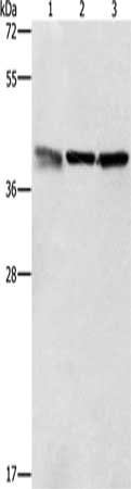
Gel: 12%SDS-PAGE, Lysate: 40 ug, Lane 1-3: A549 cells, K562 cells, PC3 cells, Primary antibody: CSB-PA238285(ZFP42 Antibody) at dilution 1/500, Secondary antibody: Goat anti rabbit IgG at 1/8000 dilution, Exposure time: 30 seconds
Gel: 12%SDS-PAGE, Lysate: 40 ug, Lane 1-3: A549 cells, K562 cells, PC3 cells, Primary antibody: CSB-PA238285(ZFP42 Antibody) at dilution 1/500, Secondary antibody: Goat anti rabbit IgG at 1/8000 dilution, Exposure time: 30 seconds
ZFP42 Antibody
CSB-PA238285
ApplicationsWestern Blot, ELISA
Product group Antibodies
ReactivityHuman
TargetZFP42
Overview
- SupplierCusabio
- Product NameZFP42 Antibody
- Delivery Days Customer20
- ApplicationsWestern Blot, ELISA
- CertificationResearch Use Only
- ClonalityPolyclonal
- ConjugateUnconjugated
- Gene ID132625
- Target nameZFP42
- Target descriptionZFP42 zinc finger protein
- Target synonymsREX-1, REX1, ZNF754, zfp-42, zinc finger protein 42 homolog, REX1 transcription factor, reduced expression protein 1, zinc finger protein 754
- HostRabbit
- IsotypeIgG
- Protein IDQ96MM3
- Protein NameZinc finger protein 42 homolog
- Scientific DescriptionRex-1 (for reduced expression), also designated zinc finger protein 42 (ZFP42), is an acidic zinc finger protein. Rex-1 contains four repeats of the zinc finger nucleic acid-binding motif and a potential acidic activator domain, suggesting that it is a regulatory protein. Rex-1 localizes to the nucleus and is highly expressed in embryonic stem (ES) and undifferentiated murine F9 teratocarcinoma cells. At the transcriptional level, expression of Rex-1 is reduced when F9 cells are induced to differentiate by the addition of retinoic acid (RA), and Rex-1 repression is enhanced by E1A. The Oct-3/4 transcription factor can either activate or repress the Rex-1 promoter, depending on the cellular environment, while Oct-6 can lower the expression of Rex-1.
- ReactivityHuman
- Storage Instruction-20°C or -80°C
- UNSPSC41116161
Related products

Product group Antibodies
Anti-Rex1/ZFP42 Antibody Picoband(r)A08177-1-CARRIER-FREE
ApplicationsFlow Cytometry, Western Blot, ELISA, ImmunoHistoChemistry
ReactivityHuman, Mouse, Rat
TargetZFP42
- SizePrice
Product group Antibodies
Zinc finger protein 754 AntibodyBS-6347R
ApplicationsImmunoFluorescence, Western Blot, ELISA, ImmunoCytoChemistry, ImmunoHistoChemistry, ImmunoHistoChemistry Frozen, ImmunoHistoChemistry Paraffin
ReactivityBovine, Equine, Human, Mouse, Porcine, Rabbit, Rat, Sheep
TargetZFP42
- SizePrice
Product group Antibodies
ZFP42 Polyclonal AntibodyCAC13463
ApplicationsImmunoFluorescence, Western Blot, ELISA
ReactivityMouse
TargetZFP42
- SizePrice
Product group Antibodies

ZFP42 / REX-1 AntibodyLS-C402920
ApplicationsWestern Blot, ELISA
ReactivityHuman
TargetZFP42
- SizePrice