

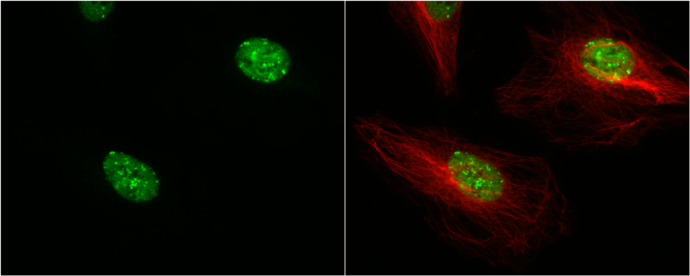
53BP1 antibody [N1], N-term detects 53BP1 protein at nucleus by immunofluorescent analysis. Sample: HeLa cells were fixed in 4% paraformaldehyde at RT for 15 min. Green: 53BP1 protein stained by 53BP1 antibody [N1], N-term (GTX102595) diluted at 1:500. Red: alpha Tubulin, a cytoskeleton marker, stained by alpha Tubulin antibody [B-5-1-2] (GTX11304) diluted at 1:10000.
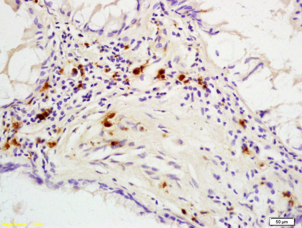
![53BP1 antibody [N1], N-term detects 53BP1 protein at nucleus by immunohistochemical analysis. Sample: Paraffin-embedded human breast carcinoma. 53BP1 stained by 53BP1 antibody [N1], N-term (GTX102595) diluted at 1:200. Antigen Retrieval: Citrate buffer, pH 6.0, 15 min](https://www.genetex.com/upload/website/prouct_img/normal/GTX102595/GTX102595_39848_20190111_IHC-P_w_23060119_607.webp "53BP1 antibody [N1], N-term detects 53BP1 protein at nucleus by immunohistochemical analysis. Sample: Paraffin-embedded human breast carcinoma. 53BP1 stained by 53BP1 antibody [N1], N-term (GTX102595) diluted at 1:200. Antigen Retrieval: Citrate buffer, pH 6.0, 15 min")
![Non-transfected (–) and transfected (+) HeLa whole cell extracts (50 μg) were separated by 5% SDS-PAGE, and the membrane was blotted with 53BP1 antibody [N1], N-term (GTX102595) diluted at 1:500.](https://www.genetex.com/upload/website/prouct_img/normal/GTX102595/GTX102595_39848_20161103_WB_shRNA_watermark_w_23060119_680.webp "Non-transfected (–) and transfected (+) HeLa whole cell extracts (50 μg) were separated by 5% SDS-PAGE, and the membrane was blotted with 53BP1 antibody [N1], N-term (GTX102595) diluted at 1:500.")
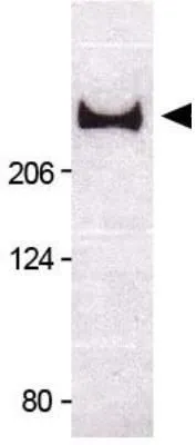
![53BP1 antibody [N1], N-term detects TP53BP1 protein by Western blot analysis. A. 30 μg 293T whole cell lysate/extract B. 30 μg HeLa whole cell lysate/extract 5 % SDS-PAGE 53BP1 antibody [N1], N-term (GTX102595) dilution: 1:500](https://www.genetex.com/upload/website/prouct_img/normal/GTX102595/GTX102595_39848_WB_w_23060119_151.webp "53BP1 antibody [N1], N-term detects TP53BP1 protein by Western blot analysis. A. 30 μg 293T whole cell lysate/extract B. 30 μg HeLa whole cell lysate/extract 5 % SDS-PAGE 53BP1 antibody [N1], N-term (GTX102595) dilution: 1:500")
![Various whole cell extracts (30 μg) were separated by 5% SDS-PAGE, and the membrane was blotted with 53BP1 antibody [N1], N-term (GTX102595) diluted at 1:500.](https://www.genetex.com/upload/website/prouct_img/normal/GTX102595/GTX102595_39848_20160128_WB_M_w_23060119_344.webp "Various whole cell extracts (30 μg) were separated by 5% SDS-PAGE, and the membrane was blotted with 53BP1 antibody [N1], N-term (GTX102595) diluted at 1:500.")
53BP1 antibody [N1], N-term detects 53BP1 protein at nucleus by immunofluorescent analysis. Sample: HeLa cells were fixed in 4% paraformaldehyde at RT for 15 min. Green: 53BP1 protein stained by 53BP1 antibody [N1], N-term (GTX102595) diluted at 1:500. Red: alpha Tubulin, a cytoskeleton marker, stained by alpha Tubulin antibody [B-5-1-2] (GTX11304) diluted at 1:10000.
53BP1 antibody [N1], N-term
GTX102595
ApplicationsImmunoFluorescence, Western Blot, ImmunoCytoChemistry, ImmunoHistoChemistry, ImmunoHistoChemistry Frozen, ImmunoHistoChemistry Paraffin
Product group Antibodies
ReactivityHuman, Mouse
TargetTP53BP1
Overview
- SupplierGeneTex
- Product Name53BP1 antibody [N1], N-term
- Delivery Days Customer9
- Application Supplier NoteWB: 1:500-1:3000. ICC/IF: 1:100-1:1000. IHC-P: 1:100-1:1000. *Optimal dilutions/concentrations should be determined by the researcher.Not tested in other applications.
- ApplicationsImmunoFluorescence, Western Blot, ImmunoCytoChemistry, ImmunoHistoChemistry, ImmunoHistoChemistry Frozen, ImmunoHistoChemistry Paraffin
- CertificationResearch Use Only
- ClonalityPolyclonal
- Concentration1 mg/ml
- ConjugateUnconjugated
- Gene ID7158
- Target nameTP53BP1
- Target descriptiontumor protein p53 binding protein 1
- Target synonyms53BP1, TDRD30, p202, p53BP1, TP53-binding protein 1, p53-binding protein 1, tumor protein 53-binding protein, 1, tumor suppressor p53-binding protein 1
- HostRabbit
- IsotypeIgG
- Protein IDQ12888
- Protein NameTP53-binding protein 1
- Scientific DescriptionMay have a role in checkpoint signaling during mitosis (By similarity). Enhances TP53-mediated transcriptional activation. Plays a role in the response to DNA damage.
- ReactivityHuman, Mouse
- Storage Instruction-20°C or -80°C,2°C to 8°C
- UNSPSC41116161
Datasheet
Related products
Product group Antibodies

Anti-TP53BP1 [RAB-C425]Ab01907-1.1
ApplicationsImmunoFluorescence, ImmunoPrecipitation
ReactivityHuman
TargetTP53BP1
- SizePrice
Product group Antibodies
Anti-53BP1 AntibodyA100698
ApplicationsELISA, ImmunoHistoChemistry
ReactivityHuman
- SizePrice
Product group Antibodies
Anti-TP53BP1 Antibody144-05757
ApplicationsImmunoFluorescence, Western Blot, ImmunoHistoChemistry
ReactivityHuman, Mouse, Rat
TargetTP53BP1
- SizePrice
Product group Antibodies

TP53BP1 / 53BP1 AntibodyLS-C663140
ApplicationsImmunoPrecipitation, Western Blot, ImmunoCytoChemistry
ReactivityHuman
TargetTP53BP1
- SizePrice

Product group Antibodies
Anti-TP53BP1 Antibody Picoband(r)A00397-1-CARRIER-FREE
ApplicationsFlow Cytometry, ImmunoFluorescence, Western Blot, ELISA, ImmunoCytoChemistry
ReactivityHuman, Mouse, Rat
TargetTP53BP1
- SizePrice
Product group Antibodies
ApplicationsImmunoPrecipitation, Western Blot, ImmunoCytoChemistry, ImmunoHistoChemistry
TargetTP53BP1
- SizePrice
Product group Antibodies
Phospho-TP53BP1 (S6) AntibodyCSB-PA050307
ApplicationsWestern Blot, ELISA, ImmunoHistoChemistry
ReactivityHuman, Monkey, Mouse, Rat
TargetTP53BP1
- SizePrice
Product group Antibodies
ApplicationsImmunoFluorescence, ELISA, ImmunoCytoChemistry, ImmunoHistoChemistry, ImmunoHistoChemistry Frozen, ImmunoHistoChemistry Paraffin
ReactivityBovine, Canine, Equine, Human, Mouse, Porcine, Rabbit, Rat
TargetTP53BP1
- SizePrice
Product group Antibodies
53BP1 antibodyGTX30658
ApplicationsImmunoFluorescence, ImmunoPrecipitation, Western Blot, ImmunoCytoChemistry, ImmunoHistoChemistry, ImmunoHistoChemistry Paraffin, Other Application
ReactivityHuman, Mouse
TargetTP53BP1
- SizePrice

Product group Antibodies
Anti-TP53BP1 AntibodyHPA008788
ApplicationsWestern Blot, ImmunoCytoChemistry
ReactivityHuman
TargetTP53BP1
- SizePrice