

ABL1 Antibody, HRP conjugated
CSB-PA04355B0RB
ApplicationsELISA
Product group Antibodies
ReactivityHuman
TargetABL1
Overview
- SupplierCusabio
- Product NameABL1 Antibody, HRP conjugated
- Delivery Days Customer20
- ApplicationsELISA
- CertificationResearch Use Only
- ClonalityPolyclonal
- ConjugateHRP
- Gene ID25
- Target nameABL1
- Target descriptionABL proto-oncogene 1, non-receptor tyrosine kinase
- Target synonymsABL, BCR-ABL, CHDSKM, JTK7, bcr/abl, c-ABL, c-ABL1, p150, v-abl, tyrosine-protein kinase ABL1, ABL protooncogene 1 nonreceptor tyrosine kinase, Abelson tyrosine-protein kinase 1, BCR-ABL1 p190, BCR/ABL e8a2 fusion, BCR/ABL1 e1a2 fusion protein, BCR/ABL1 fusion, BCR/ABL1 fusion protein e3a1, BCR::ABL1 fusion protein, bcr/c-abl oncogene protein, c-abl oncogene 1, receptor tyrosine kinase, chimeric BCR::ABL1 protein, proto-oncogene c-Abl, proto-oncogene tyrosine-protein kinase ABL1, v-abl Abelson murine leukemia viral oncogene homolog 1
- HostRabbit
- IsotypeIgG
- Protein IDP00519
- Protein NameTyrosine-protein kinase ABL1
- Scientific DescriptionNon-receptor tyrosine-protein kinase that plays a role in many key processes linked to cell growth and survival such as cytoskeleton remodeling in response to extracellular stimuli, cell motility and adhesion, receptor endocytosis, autophagy, DNA damage response and apoptosis. Coordinates actin remodeling through tyrosine phosphorylation of proteins controlling cytoskeleton dynamics like WASF3 (involved in branch formation); ANXA1 (involved in membrane anchoring); DBN1, DBNL, CTTN, RAPH1 and ENAH (involved in signaling); or MAPT and PXN (microtubule-binding proteins). Phosphorylation of WASF3 is critical for the stimulation of lamellipodia formation and cell migration. Involved in the regulation of cell adhesion and motility through phosphorylation of key regulators of these processes such as BCAR1, CRK, CRKL, DOK1, EFS or NEDD9. Phosphorylates multiple receptor tyrosine kinases and more particularly promotes endocytosis of EGFR, facilitates the formation of neuromuscular synapses through MUSK, inhibits PDGFRB-mediated chemotaxis and modulates the endocytosis of activated B-cell receptor complexes. Other substrates which are involved in endocytosis regulation are the caveolin (CAV1) and RIN1. Moreover, ABL1 regulates the CBL family of ubiquitin ligases that drive receptor down-regulation and actin remodeling. Phosphorylation of CBL leads to increased EGFR stability. Involved in late-stage autophagy by regulating positively the trafficking and function of lysosomal components. ABL1 targets to mitochondria in response to oxidative stress and thereby mediates mitochondrial dysfunction and cell death. In response to oxidative stress, phosphorylates serine/threonine kinase PRKD2 at Tyr-717 (PubMed:28428613). ABL1 is also translocated in the nucleus where it has DNA-binding activity and is involved in DNA-damage response and apoptosis. Many substrates are known mediators of DNA repair: DDB1, DDB2, ERCC3, ERCC6, RAD9A, RAD51, RAD52 or WRN. Activates the proapoptotic pathway when the DNA damage is too severe to be repaired. Phosphorylates TP73, a primary regulator for this type of damage-induced apoptosis. Phosphorylates the caspase CASP9 on Tyr-153 and regulates its processing in the apoptotic response to DNA damage. Phosphorylates PSMA7 that leads to an inhibition of proteasomal activity and cell cycle transition blocks. ABL1 acts also as a regulator of multiple pathological signaling cascades during infection. Several known tyrosine-phosphorylated microbial proteins have been identified as ABL1 substrates. This is the case of A36R of Vaccinia virus, Tir (translocated intimin receptor) of pathogenic E.coli and possibly Citrobacter, CagA (cytotoxin-associated gene A) of H.pylori, or AnkA (ankyrin repeat-containing protein A) of A.phagocytophilum. Pathogens can highjack ABL1 kinase signaling to reorganize the host actin cytoskeleton for multiple purposes, like facilitating intracellular movement and host cell exit. Finally, functions as its own regulator through autocatalytic activity as well as through phosphorylation of its inhibitor, ABI1. Regulates T-cell differentiation in a TBX21-dependent manner. Phosphorylates TBX21 on tyrosine residues leading to an enhancement of its transcriptional activator activity (By similarity).
- ReactivityHuman
- Storage Instruction-20°C or -80°C
- UNSPSC41116161
Related products
Product group Antibodies
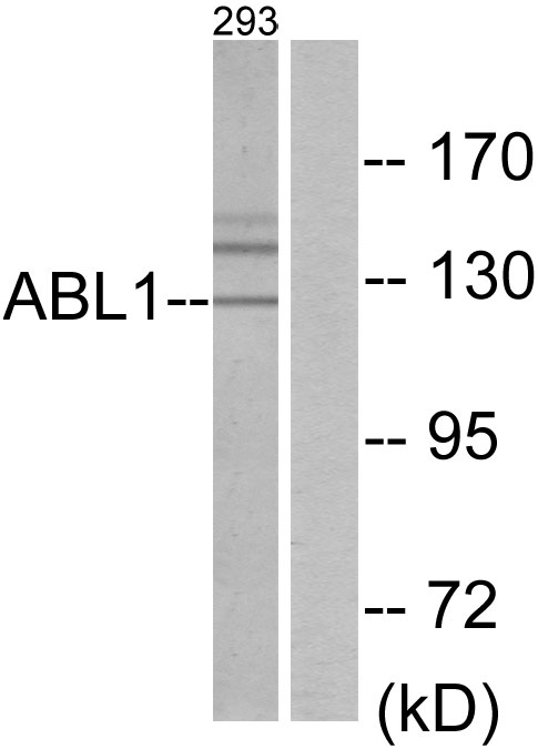
Anti-ABL1 Antibody144-00282
ApplicationsImmunoFluorescence, Western Blot, ImmunoHistoChemistry
ReactivityHuman, Mouse, Rat
TargetABL1
- SizePrice

Product group Antibodies
Anti-ABL1 Antibody Picoband(r)A00133-1-CARRIER-FREE
ApplicationsFlow Cytometry, ImmunoFluorescence, Western Blot, ELISA, ImmunoCytoChemistry
ReactivityHuman, Mouse, Rat
TargetABL1
- SizePrice
Product group Antibodies
References
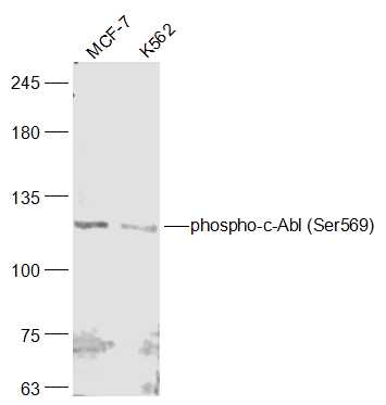
c-Abl antibodyGTX111317
ApplicationsImmunoFluorescence, Western Blot, ImmunoCytoChemistry, ImmunoHistoChemistry, ImmunoHistoChemistry Paraffin
ReactivityHuman
TargetABL1
- SizePrice
Product group Antibodies
ABL1 Polyclonal AntibodyCAC14060
ApplicationsImmunoPrecipitation, ELISA, ImmunoHistoChemistry
TargetABL1
- SizePrice
Product group Antibodies
ApplicationsImmunoFluorescence, Western Blot, ELISA, ImmunoCytoChemistry, ImmunoHistoChemistry, ImmunoHistoChemistry Frozen, ImmunoHistoChemistry Paraffin
ReactivityBovine, Equine, Human, Porcine, Sheep
TargetABL1
- SizePrice
Product group Antibodies
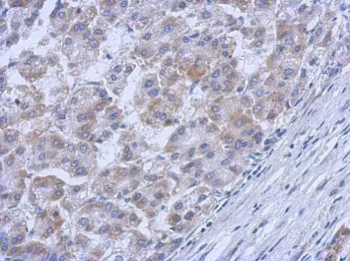
ABL1 AntibodyCSB-PA001140
ApplicationsWestern Blot, ELISA, ImmunoHistoChemistry
ReactivityHuman, Monkey, Mouse, Rat
TargetABL1
- SizePrice
Product group Antibodies
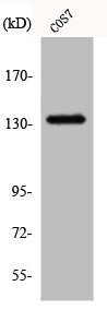
Anti-ABL1 AntibodyA96250
ApplicationsWestern Blot, ELISA, ImmunoHistoChemistry
ReactivityHuman, Mouse, Rat
- SizePrice

Product group Antibodies
Anti-ABL1 AntibodyHPA027251
ApplicationsImmunoHistoChemistry
ReactivityHuman
TargetABL1
- SizePrice