

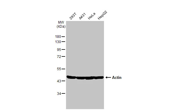
Various whole cell extracts (30 μg) were separated by 10% SDS-PAGE, and the membrane was blotted with Actin antibody (GTX638580) diluted at 1:10000. The HRP-conjugated anti-rabbit IgG antibody (GTX213110-01) was used to detect the primary antibody.
![Whole cell extract (30 μg) was separated by 10% SDS-PAGE, and the membrane was blotted with Actin antibody [HL2372] (GTX638580) diluted at 1:50000. The HRP-conjugated anti-rabbit IgG antibody (GTX213110-01) was used to detect the primary antibody.](https://www.genetex.com/upload/website/prouct_img/normal/GTX638580/GTX638580_T-45040_20230616_WB_R_23062019_652.webp "Whole cell extract (30 μg) was separated by 10% SDS-PAGE, and the membrane was blotted with Actin antibody [HL2372] (GTX638580) diluted at 1:50000. The HRP-conjugated anti-rabbit IgG antibody (GTX213110-01) was used to detect the primary antibody.")
![Whole cell extract (30 μg) was separated by 10% SDS-PAGE, and the membrane was blotted with Actin antibody [HL2372] (GTX638580) diluted at 1:50000. The HRP-conjugated anti-rabbit IgG antibody (GTX213110-01) was used to detect the primary antibody.](https://www.genetex.com/upload/website/prouct_img/normal/GTX638580/GTX638580_T-45040_20230616_WB_R_2_23062019_494.webp "Whole cell extract (30 μg) was separated by 10% SDS-PAGE, and the membrane was blotted with Actin antibody [HL2372] (GTX638580) diluted at 1:50000. The HRP-conjugated anti-rabbit IgG antibody (GTX213110-01) was used to detect the primary antibody.")
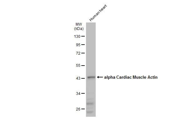
![Various whole cell extracts (30 μg) were separated by 10% SDS-PAGE, and the membrane was blotted with Actin antibody [HL2372] (GTX638580) diluted at 1:10000. The HRP-conjugated anti-rabbit IgG antibody (GTX213110-01) was used to detect the primary antibody.](https://www.genetex.com/upload/website/prouct_img/normal/GTX638580/GTX638580_T-45040_20230616_WB_M_23062019_191.webp "Various whole cell extracts (30 μg) were separated by 10% SDS-PAGE, and the membrane was blotted with Actin antibody [HL2372] (GTX638580) diluted at 1:10000. The HRP-conjugated anti-rabbit IgG antibody (GTX213110-01) was used to detect the primary antibody.")
![Actin antibody [HL2372] detects Actin protein at cytoskeleton by immunofluorescent analysis.
Sample: HeLa cells were fixed in ice-cold MeOH for 5 min.
Green: Actin stained by Actin antibody [HL2372] (GTX638580) diluted at 1:1000.
Blue: Fluoroshield with DAPI (GTX30920).](https://www.genetex.com/upload/website/prouct_img/normal/GTX638580/GTX638580_T-45040_20230609_ICC_IF_23062718_886.webp "Actin antibody [HL2372] detects Actin protein at cytoskeleton by immunofluorescent analysis.
Sample: HeLa cells were fixed in ice-cold MeOH for 5 min.
Green: Actin stained by Actin antibody [HL2372] (GTX638580) diluted at 1:1000.
Blue: Fluoroshield with DAPI (GTX30920).")
![Various whole cell extracts were separated by 10% SDS-PAGE, and the membrane was blotted with Actin antibody [HL2372] (GTX638580) diluted at 1:10000. The HRP-conjugated anti-rabbit IgG antibody (GTX213110-01) was used to detect the primary antibody.](https://www.genetex.com/upload/website/prouct_img/normal/GTX638580/GTX638580_45131_20230901_WB_Sensitivity_23090619_569.webp "Various whole cell extracts were separated by 10% SDS-PAGE, and the membrane was blotted with Actin antibody [HL2372] (GTX638580) diluted at 1:10000. The HRP-conjugated anti-rabbit IgG antibody (GTX213110-01) was used to detect the primary antibody.")
![Actin antibody [HL2372] detects Actin protein by immunohistochemical analysis.
Sample: Paraffin-embedded mouse tissues.
Actin stained by Actin antibody [HL2372] (GTX638580) diluted at 1:100.
Antigen Retrieval: Citrate buffer, pH 6.0, 15 min](https://www.genetex.com/upload/website/prouct_img/normal/GTX638580/GTX638580_T-45040_20230928_IHC-P_multiple_M_23102401_404.webp "Actin antibody [HL2372] detects Actin protein by immunohistochemical analysis.
Sample: Paraffin-embedded mouse tissues.
Actin stained by Actin antibody [HL2372] (GTX638580) diluted at 1:100.
Antigen Retrieval: Citrate buffer, pH 6.0, 15 min")
![Actin antibody [HL2372] detects Actin protein by immunohistochemical analysis.
Sample: Paraffin-embedded rat tissues.
Actin stained by Actin antibody [HL2372] (GTX638580) diluted at 1:100.
Antigen Retrieval: Citrate buffer, pH 6.0, 15 min](https://www.genetex.com/upload/website/prouct_img/normal/GTX638580/GTX638580_T-45040_20230928_IHC-P_multiple_R_23102401_191.webp "Actin antibody [HL2372] detects Actin protein by immunohistochemical analysis.
Sample: Paraffin-embedded rat tissues.
Actin stained by Actin antibody [HL2372] (GTX638580) diluted at 1:100.
Antigen Retrieval: Citrate buffer, pH 6.0, 15 min")
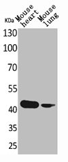
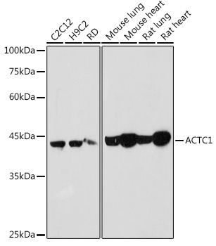
![Various whole cell extracts (30 μg) were separated by 10% SDS-PAGE, and the membrane was blotted with Actin antibody [HL2372] (GTX638580) diluted at 1:10000. The HRP-conjugated anti-rabbit IgG antibody (GTX213110-01) was used to detect the primary antibody.](https://www.genetex.com/upload/website/prouct_img/normal/GTX638580/GTX638580_45131_20231110_WB_multiple_species_23111422_691.webp "Various whole cell extracts (30 μg) were separated by 10% SDS-PAGE, and the membrane was blotted with Actin antibody [HL2372] (GTX638580) diluted at 1:10000. The HRP-conjugated anti-rabbit IgG antibody (GTX213110-01) was used to detect the primary antibody.")
Various whole cell extracts (30 μg) were separated by 10% SDS-PAGE, and the membrane was blotted with Actin antibody (GTX638580) diluted at 1:10000. The HRP-conjugated anti-rabbit IgG antibody (GTX213110-01) was used to detect the primary antibody.
Actin antibody [HL2372]
GTX638580
ApplicationsImmunoFluorescence, Western Blot, ImmunoCytoChemistry, ImmunoHistoChemistry, ImmunoHistoChemistry Paraffin
Product group Antibodies
ReactivityCanine, Drosophila, Feline, Human, Monkey, Mouse, Rabbit, Rat
TargetACTC1
Overview
- SupplierGeneTex
- Product NameActin antibody [HL2372]
- Delivery Days Customer9
- Application Supplier NoteWB: 1:1000-1:10000. *Optimal dilutions/concentrations should be determined by the researcher.Not tested in other applications.
- ApplicationsImmunoFluorescence, Western Blot, ImmunoCytoChemistry, ImmunoHistoChemistry, ImmunoHistoChemistry Paraffin
- CertificationResearch Use Only
- ClonalityMonoclonal
- Clone IDHL2372
- Concentration1 mg/ml
- ConjugateUnconjugated
- Gene ID70
- Target nameACTC1
- Target descriptionactin alpha cardiac muscle 1
- Target synonymsACTC, ASD5, CMD1R, CMH11, LVNC4, actin, alpha cardiac muscle 1
- HostRabbit
- IsotypeIgG
- Protein IDP68032
- Protein NameActin, alpha cardiac muscle 1
- Scientific DescriptionActins are highly conserved proteins that are involved in various types of cell motility. Polymerization of globular actin (G-actin) leads to a structural filament (F-actin) in the form of a two-stranded helix. Each actin can bind to four others. The protein encoded by this gene belongs to the actin family which is comprised of three main groups of actin isoforms, alpha, beta, and gamma. The alpha actins are found in muscle tissues and are a major constituent of the contractile apparatus. Defects in this gene have been associated with idiopathic dilated cardiomyopathy (IDC) and familial hypertrophic cardiomyopathy (FHC). [provided by RefSeq, Jul 2008]
- ReactivityCanine, Drosophila, Feline, Human, Monkey, Mouse, Rabbit, Rat
- Storage Instruction-20°C or -80°C,2°C to 8°C
- UNSPSC12352203
Datasheet
Related products
Product group Antibodies
ApplicationsWestern Blot, ImmunoHistoChemistry, ImmunoHistoChemistry Paraffin
ReactivityHuman, Mouse
TargetACTC1
- SizePrice
Product group Antibodies
References
ApplicationsFlow Cytometry, ImmunoFluorescence, Western Blot, ELISA, ImmunoCytoChemistry, ImmunoHistoChemistry, ImmunoHistoChemistry Frozen, ImmunoHistoChemistry Paraffin
ReactivityHuman, Mouse, Rabbit, Rat
TargetACTC1
- SizePrice
Product group Antibodies
ACTC1 AntibodyCSB-PA004766
ApplicationsWestern Blot, ELISA
ReactivityHuman, Mouse, Rat
TargetACTC1
- SizePrice
Product group Antibodies
ApplicationsImmunoPrecipitation, Western Blot, ImmunoCytoChemistry, ImmunoHistoChemistry
ReactivityBovine, Canine, Chicken, Equine, Guinea Pig, Mouse, Porcine, Rat
TargetACTC1
- SizePrice
Product group Antibodies
Actin antibodyGTX14130
ApplicationsELISA, ImmunoHistoChemistry, ImmunoHistoChemistry Frozen, ImmunoHistoChemistry Paraffin
ReactivityChicken, Human
- SizePrice
Product group Antibodies

Mouse anti actin alpha-cardiacMUB0109P
ApplicationsWestern Blot, ELISA, ImmunoHistoChemistry, ImmunoHistoChemistry Frozen, ImmunoHistoChemistry Paraffin
ReactivityHuman, Mammals, Porcine, Rabbit, Rat, Zebra Fish
TargetACTC1
- SizePrice
Product group Antibodies
References
alpha Cardiac Muscle Actin antibodyGTX101876
ApplicationsImmunoFluorescence, Western Blot, ImmunoCytoChemistry, ImmunoHistoChemistry, ImmunoHistoChemistry Paraffin
ReactivityHuman, Mouse, Rat
TargetACTC1
- SizePrice
![ICC/IF analysis of 3T3 cells using GTX80809 Actin antibody [mAbGEa]. Green : Primary antibody Blue : cell nucleus Permeabilization : 0.1% Triton X-100 in TBS for 15 minutes at room temperature Dilution : 1:100 in blocking buffer for at least 1 hour at room temperature](https://www.genetex.com/upload/website/prouct_img/normal/GTX80809/GTX80809_904_ICC-IF_w_23061322_169.webp)
Product group Antibodies
References
Actin antibody [mAbGEa]GTX80809
ApplicationsImmunoFluorescence, ImmunoPrecipitation, Western Blot, ChIP Chromatin ImmunoPrecipitation, ELISA, ImmunoCytoChemistry, ImmunoHistoChemistry, ImmunoHistoChemistry Paraffin
ReactivityBacteria, Bovine, Drosophila, Human, Mouse, Plant, Porcine, Rat, Sheep, Xenopus, Yeast, Zebra Fish
TargetACTC1
- SizePrice
Product group Antibodies
Anti-ACTC1 AntibodyA80440
ApplicationsWestern Blot, ImmunoHistoChemistry
ReactivityHuman, Mouse, Rat
- SizePrice