

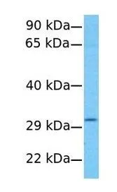
WB analysis of HeLa cells using GTX46672 AGPAT2 antibody at 1.0μg/ml.

WB analysis of HeLa cells using GTX46672 AGPAT2 antibody at 1.0μg/ml.
AGPAT2 antibody, C-term
GTX46672
ApplicationsWestern Blot
Product group Antibodies
ReactivityHuman
TargetAGPAT2
Overview
- SupplierGeneTex
- Product NameAGPAT2 antibody, C-term
- Delivery Days Customer9
- ApplicationsWestern Blot
- CertificationResearch Use Only
- ClonalityPolyclonal
- Concentration0.5-1 mg/ml
- ConjugateUnconjugated
- Gene ID10555
- Target nameAGPAT2
- Target description1-acylglycerol-3-phosphate O-acyltransferase 2
- Target synonyms1-AGPAT2, BSCL, BSCL1, LPAAB, LPAAT-beta, LPLAT2, 1-acyl-sn-glycerol-3-phosphate acyltransferase beta, 1-AGP acyltransferase 2, 1-acylglycerol-3-phosphate O-acyltransferase 2 (lysophosphatidic acid acyltransferase, beta), lysophosphatidic acid acyltransferase-beta, lysophospholipid acyltransferase 2, testicular tissue protein Li 143
- HostRabbit
- IsotypeIgG
- Protein IDO15120
- Protein Name1-acyl-sn-glycerol-3-phosphate acyltransferase beta
- Scientific DescriptionThis gene encodes a member of the 1-acylglycerol-3-phosphate O-acyltransferase family. The protein is located within the endoplasmic reticulum membrane and converts lysophosphatidic acid to phosphatidic acid, the second step in de novo phospholipid biosynthesis. Mutations in this gene have been associated with congenital generalized lipodystrophy (CGL), or Berardinelli-Seip syndrome, a disease characterized by a near absence of adipose tissue and severe insulin resistance. Alternate transcriptional splice variants, encoding different isoforms, have been characterized. [provided by RefSeq, Jul 2008]
- ReactivityHuman
- Storage Instruction-20°C or -80°C,2°C to 8°C
- UNSPSC41116161
Datasheet
Related products

Product group Antibodies
Anti-AGPAT2 Antibody Picoband(r)A04744-1-CARRIER-FREE
ApplicationsWestern Blot, ELISA, ImmunoHistoChemistry
ReactivityHuman, Mouse, Rat
TargetAGPAT2
- SizePrice
Product group Antibodies
Anti-AGPAT2 AntibodyA29556
ApplicationsImmunoFluorescence, Western Blot, ImmunoHistoChemistry
ReactivityHuman
- SizePrice
Product group Antibodies
Anti-AGPAT2 Antibody144-66167
ApplicationsImmunoFluorescence, Western Blot
ReactivityHuman, Mouse
TargetAGPAT2
- SizePrice
Product group Antibodies
Agpat2 Polyclonal AntibodyBS-5032R
ApplicationsImmunoFluorescence, ELISA, ImmunoCytoChemistry, ImmunoHistoChemistry, ImmunoHistoChemistry Frozen, ImmunoHistoChemistry Paraffin
ReactivityBovine, Canine, Equine, Human, Mouse, Porcine, Rat
TargetAGPAT2
- SizePrice
Product group Antibodies
AGPAT2 AntibodyCSB-PA001450ESR2HU
ApplicationsWestern Blot, ELISA, ImmunoHistoChemistry
ReactivityHuman
TargetAGPAT2
- SizePrice
Product group Antibodies
Agpat2 Polyclonal AntibodyCAC10713
ApplicationsWestern Blot, ELISA, ImmunoHistoChemistry
TargetAGPAT2
- SizePrice
Product group Antibodies

AGPAT2 AntibodyLS-C402605
ApplicationsWestern Blot, ELISA
ReactivityHuman
TargetAGPAT2
- SizePrice

Product group Antibodies
Anti-AGPAT2 AntibodyHPA019544
ApplicationsWestern Blot, ImmunoHistoChemistry
ReactivityHuman
TargetAGPAT2
- SizePrice
Product group Antibodies
Anti-AGPAT2Y158051
ApplicationsELISA, ImmunoHistoChemistry
ReactivityHuman, Mouse, Rat
- SizePrice